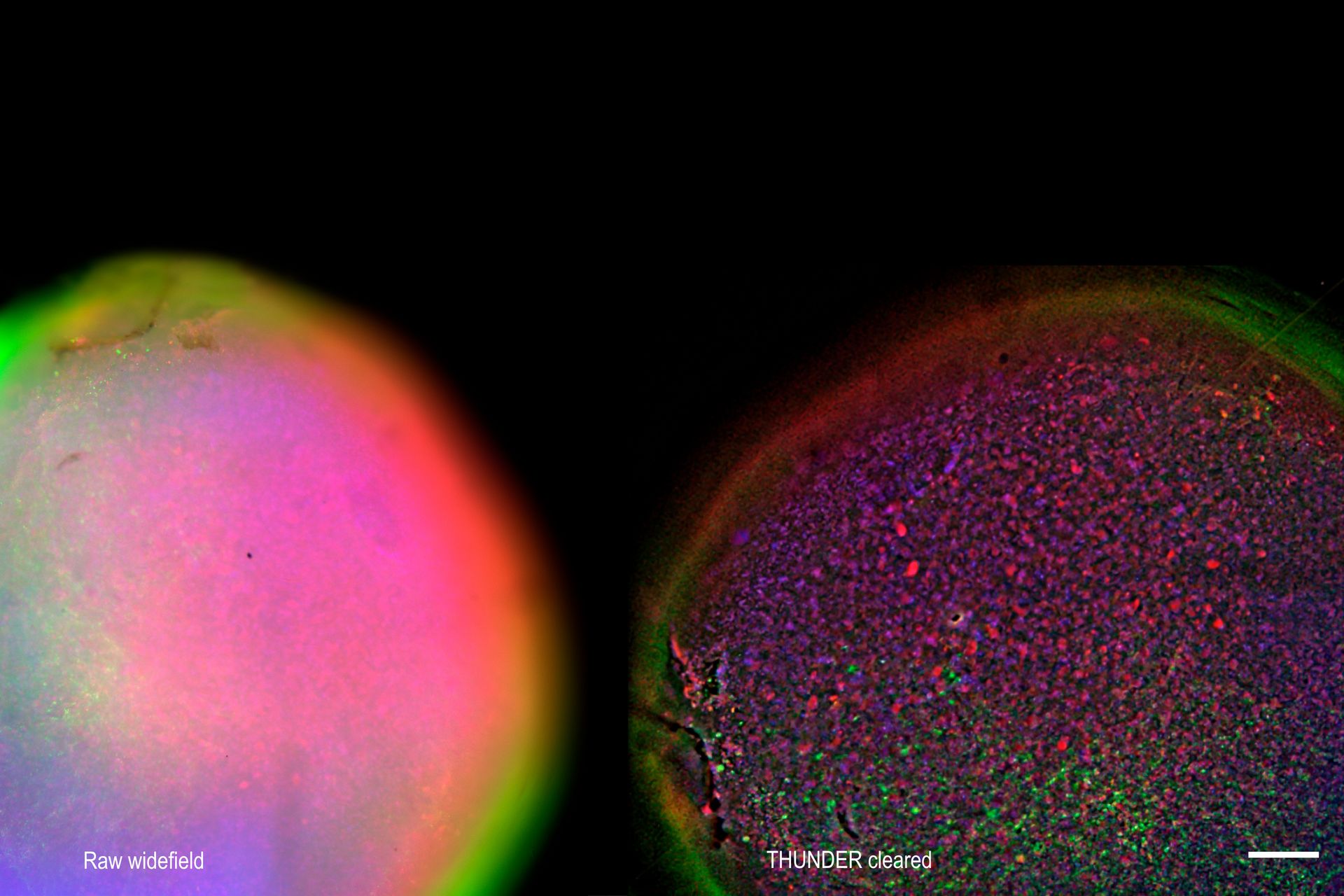
 2D slices of a 1 mm diameter midbrain neural organoid stained with DAPI (blue, nuclear stain), β-tubulin (green, neuronal stain), and GFAP (red, astrocyte stain).")
Introduction
Live‑cell imaging of large, complex biological samples often requires large fields of view, sub-cellular resolution, high-sensitivity, and fast acquisition – all while maintaining low illumination doses for gentle long-term observation. Widefield fluorescence microscopy can meet these demands, but signals can be buried in its inherent out of focus blur. Optical sectioning microscopy techniques can mitigate this, but can introduce trade-offs in speed, phototoxicity, or experimental complexity.
How can you achieve high-speed, high‑contrast imaging of large and optically challenging samples with low phototoxicity?
Widefield microscopy remains a powerful approach for live-cell imaging—provided that background fluorescence is effectively suppressed. This application note demonstrates how THUNDER Imager Cell, paired with the Teledyne Photometrics Kinetix22 sCMOS camera, delivers a balanced, live cell friendly imaging solution for a broad range of discovery biology applications. After outlining how to optimize imaging parameters for live-cell experiments, we highlight performance across diverse sample types.
Case studies include:
- 2D imaging of large field of view (FOV) U2OS and HeLa cells
- Imaging of neural organoids
- Live imaging of zebrafish embryos
 2D slices of a 1 mm diameter midbrain neural organoid stained with DAPI (blue, nuclear stain), β-tubulin (green, neuronal stain), and GFAP (red, astrocyte stain).")
 and phalloidin (magenta), imaged using Viventis SCAPE; scale bar 50μm. Courtesy of Marina Cuenca and Heleen Jungen (Dayton lab), EMBL Barcelona.")