
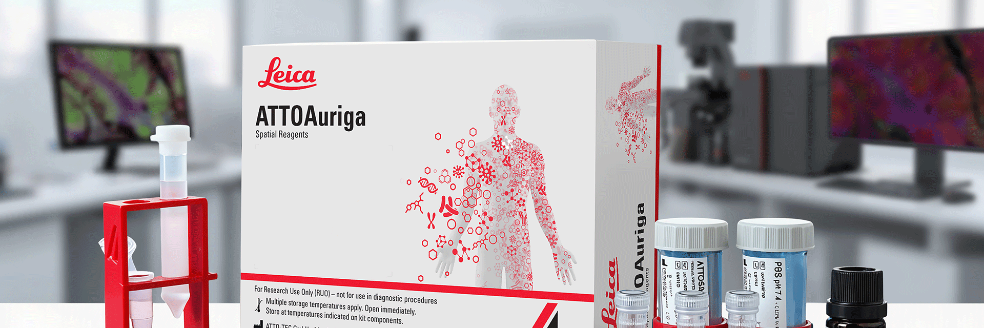
Spatial Biology Reagents
Spatial biology researchers face increasing pressure to extract deeper biological insight from limited tissue. Traditional multiplex immunofluorescence workflows can be time-consuming and difficult to reproduce, often requiring trade-offs between plex level, signal quality, and experimental confidence.
ATTOAuriga, a modular spatial biology reagent ecosystem
Introducing ATTOAuriga, a modular spatial biology reagent ecosystem that enables researchers to build and adapt multiplex experiments using a defined set of interoperable reagents, from pre-validated antibody panels to conjugation kits for custom antibody barcoding, supporting confident cyclic imaging workflows.
Contact a local imaging specialist for expert advice on the right solution for your needs and budget.
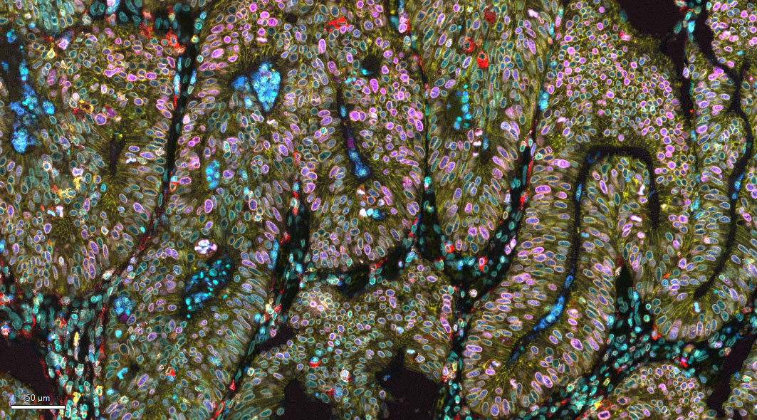
Key Benefits

Bright, clean signals
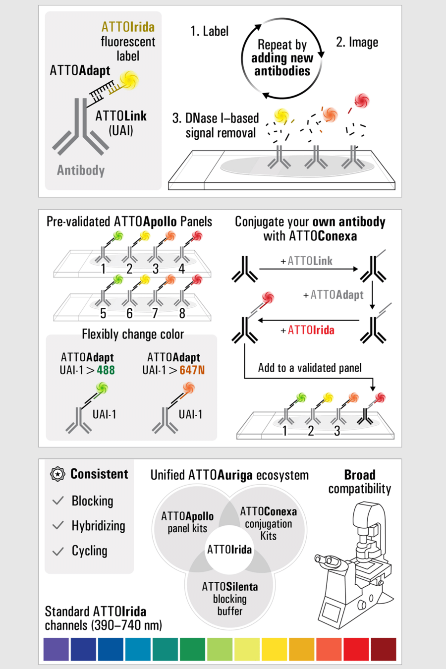
- Uses ATTOIrida fluorescent labels that hybridize specifically to Unique Antibody Identifiers (UAI) on antibodies
- Enzyme (DNase I) based signal removal clears fluorescence between cycles, minimizing signal carryover
- Designed from the ground-up for advanced high-plex immunofluorescence imaging

Built-in flexibility
- Pre-validated ATTOApollo panels for ready-to-use workflows
- ATTOConexa kits enable users to barcode their own antibodies
- Antibody barcoding allows flexible mapping of fluorescent labels through ATTOAdapt adapters
- Supports integration of user-generated antibodies alongside validated panels

Designed for reproducibility
- Defined reagent ecosystem with standardized components
- Validated for human FFPE tissue sections
- Consistent blocking, hybridization, and cycling chemistry across experiment
- Suitable for fluorescence microscopes with detection spectra compatible with ATTOIrida labels
A modular approach to multiplex imaging
Building multiplex antibody panels can be time-consuming and costly, which can slow progress and increase experimental risk for spatial biology researchers.
ATTOAuriga, a modular reagent family
ATTOAuriga is a modular reagent family that supports repeated rounds of antibody labeling, imaging, and enzymatic signal removal. Each component plays a defined role in delivering reliable cyclic imaging performance while maintaining tissue integrity and signal quality.
- ATTOIrida fluorescent labels that hybridize specifically to Unique Antibody Identifiers (UAI) on antibodies
- Pre-validated ATTOApollo panels for ready-to-use workflows
- Defined reagent ecosystem with standardized components
Curious how ATTOAuriga can fit into your workflow?
Contact us to discuss panels, conjugation options, and cyclic imaging strategies.
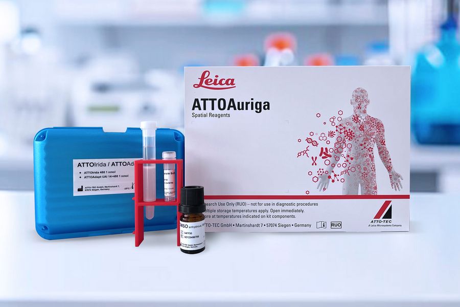
Each component has a distinct role
ATTOApollo Panels
Pre-validated, ready-to-use antibody panels with Unique
Antibody Identifiers (UAI) barcodes.
ATTOConexa Kits
Conjugation kits for generating oligonucleotide barcoded
antibodies.
ATTOIrida Fluorescent Labels
Hybridization-based labels designed for cyclic imaging
workflows.
ATTOSilenta Blocking Buffer
Required buffer optimized for signal quality and reproducibility.
Note: This material was created and approved prior to the product launch, but only as a pre-announcement.
Spatial Biology Reagents FAQs
Antibodies are added to the sample for an incubation at room temperature. After antibody binding, ATTOAdapt molecules particular to the antibodies
ATTOApollo kits may be freely combined to build large multiplex panels. If building custom panels using ATTOConexa conjugates, each conjugate must receive a unique UAI and ATTOAdapt pair. Currently, we can support up to eight 4 color multiplexed imaging rounds in the 488, 550, 647N, and 740H configuration. For other color configurations, we can support 2 rounds using any of the available ATTOIrida colors.
Presently, ATTOAuriga reagents have been validated for use with thin (2-5 micron thick) sections. Thicker sections may require specialized protocols for effective staining.
Each ATTOApollo kit can label 20 slides using a 150 microliter reaction volume.
Tissue should generally be sectioned to 2 to 5 microns in thickness. Specific tissue preparation guidelines for downstream staining workflows should be observed.
Material data safety sheets can be found here. ATTOAuriga reagents use DNA oligonucleotide technology, and so DNase free plastics, reagents, and laboratory best practices should be used.
Please contact our spatial biology Customer Success team.
Yes. By removing DNA oligos from ATTOApollo or ATTOConexa conjugates following imaging using DNAseI, dye molecules can be removed from the sample and subsequent relabeling and imaging can be performed. When generating custom antibody conjugates using the ATTOConexa kit, ensure that unique UAIs are used for each conjugate in the experiment and that the ATTOAdapt sequences are properly configured to bind the correct dyes in each multiplexing round.
No specialized mounting media is required for the ATTOAuriga workflow. Slides should be imaged in PBS under a Type 1.5 coverslip, as described in the protocol. Conventional antifade or hard‑set mounting media (e.g., ProLong Gold, Fluoromount, Vectashield) should not be used, because they will interfere with cyclic imaging, labeling, and erasure steps.
If permanent mounting is desired after the final imaging cycle, users may optionally apply a standard antifade mounting medium at the end of the workflow.
When using a Cell DIVE system, slides can be coverslipped or mounted in a ClickWell following the standard Cell DIVE protocol and using the Standard Coverslipped or ClickWell mounting media respectively.
Cell DIVE software should be updated to at least version 5.0.0.11 to facilitate the panel selection options available for ATTOApollo panels. Contact ASLS (babsupport@advancedsolutions.com) to acquire the latest ATTOAuriga BioApp version if using the BAB200 robotics system.
To integrate ATTOApollo into the Cell DIVE workflow, separate ATTOApollo conjugated antibodies into their own Cell DIVE rounds and process those rounds following the ATTOApollo staining protocol and DNaseI erasure method. Slides should follow the ATTOApollo ER2 antigen retrieval process during slide preparation. To automate ATTOApollo rounds on the BAB200 robotics system, use the ATTOAuriga BioApp.
Slides may be stored in PBS at 4 °C for up to one day during PBS wash steps.
Yes. DAPI must be reapplied in every cycle because repeated washing, labeling, and signal‑erasure steps reduce nuclear signal, and restaining mitigates this loss.
No purification is required. Excess ATTOLink does not interfere with staining.
The workflow supports multiple rounds of staining, imaging, and DNase‑based signal removal.
ATTOSilenta blocking buffer is required. It improves signal‑to‑background and is fully compatible with hybridization and DNase I cycling.
The ATTOApollo staining protocol can be automated on a BAB200 robotics system using the ATTOAuriga BioApp. BAB200 users can contact ASLS (babsupport@advancedsolutions.com) to receive the BAB200 software update.
Any fluorescence microscope capable of detecting ATTOIrida channels (e.g., 488, 550, 647, 740 nm). Use type 1.5 coverslips and image in PBS as described.
After antigen retrieval, slides can be stored in PBS at 4 °C for several days prior to staining.
For longer term storage, slides should be coverslipped with 90% glycerol mounting media with antifade reagent and stored upright in a dark contained at 4 °C.
A final cycle of directly dye-conjugated antibodies may be included following ATTOApollo staining rounds. If additional rounds are desired, the dyes bound to these antibodies will need to be removed.
Yes. Users may barcode their own antibodies using ATTOConexa kits. We advise users that 40-60% of IgG monoclonal antibodies or recombinant antibodies are expected to be compatible with ATTOConexa oligonucleotide conjugation. Users are advised to check our ATTOConexa compatibility list for clones that have been confirmed to be ATTOConexa-compatible. Once barcoded, antibodies can be integrated with ATTOApollo panels. For secondary‑based workflows, please contact technical support (contact details).
Compatible antibodies must be carrier‑free, BSA‑free, azide‑free IgG in an amine‑free buffer at ~0.2–1 mg/mL. Recommended starting amount: 100 µg.
ATTOApollo panels are optimized for the following antigen retrieval conditions: Leica Bond ER2 Buffer (pH 9.0) with heat‑mediated antigen retrieval at 95 °C for 30 minutes, followed by cooling and PBS washes. Users may explore alternative methods, but these would require independent validation, as deviations from the ER2 protocol may impact staining quality or cyclic imaging performance.
The ATTOAuriga workflow requires users to supply DNase I, standard FFPE slides, deparaffinization and antigen‑retrieval equipment, a humidity chamber for labeling steps, type 1.5 coverslips for imaging, DAPI (e.g., example), and routine lab consumables needed for washing, handling, and mounting tissue sections.
ATTOApollo Panels are validated specifically for human FFPE tissue sections. Other sample types have not been validated.
The ATTOAuriga ecosystem is a reagent family that enables cyclic immunofluorescence using DNA-barcoded antibodies. Each component has a distinct role:
- ATTOApollo (Pre-validated Antibody Panels) Panels contain ready-to-use validated antibodies labeled with ATTOLink UAI (Unique Antibody Identifier) barcodes. Each kit contains ATTOIrida fluorescent labels, ATTOAdapt adapters, and ATTOSilenta blocking buffer. These antibodies are fully validated for use in the ATTOAuriga cyclic imaging workflow.
- ATTOConexa Kits generate UAI barcoded antibodies, matching the same UAI system used by ATTOApollo Panels. They include a UAI containing ATTOLink barcode and an ATTOAdapt connector oligonucleotide, enabling flexible mapping of UAIs to specific ATTOIrida labels.
- ATTOIrida are next generation fluorescent labels, which are available in 13 different colors covering 390nm to 740nm spectral range. ATTOIrida labels comprise a color-specific DNA-barcode that allows their attachment to barcoded affinity reagents like ATTOApollo or ATTOConexa antibodies via ATTOAdapt adapters, which map a UAI to a color-specific barcode. This enables to flexibly change the color assignment for an ATTOApollo or ATTOConxea marker using a different ATTOAdapt adapter. ATTOIrida labels may also be attached in the same way to other suitably barcoded reagents such as (FISH probes for RNA detection, or microspheres).
- ATTOAdapt (UAI Adapter Oligo) is an adapter oligonucleotide used in UAI workflows. It binds the UAI barcode on the antibody and “adapts” it to a specific ATTOIrida fluorescent label. It is included in ATTOApollo Panels and ATTOConexa Kits.
- ATTOLink (Barcode Oligonucleotide) provides the UAI barcode that is chemically conjugated to each antibody.
- ATTOSilenta (Blocking Buffer) is the required blocking buffer designed for use with ATTOApollo panels, ATTOConexa kits, and ATTOIrida fluorescent labels. It reduces nonspecific binding and is an essential part of every staining cycle.


