연락처
연락처

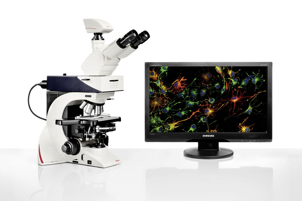
Leica DM2500 & DM2500 LED 강력한 광학 성능의 최첨단 인체공학적 현미경 시스템

최신 기사를 읽어 보세요

Staining is crucial to revealing details in the specimens.

H&E Staining in Microscopy

If we consider the role of microscopy in pathologists’ daily routines, we often think of the diagnosis. While microscopes indeed play a crucial role at this stage of the pathology lab workflow, they…
Acute lymphoblastic leukemia

How to Benefit from Digital Cytopathology

If you have thought of digital cytopathology as characterized by the digitization of glass slides, this webinar with Dr. Alessandro Caputo from the University Hospital of Salerno, Italy will broaden…
Laboratory

Factors for Selecting Clinical Microscopes

If you are considering the purchase of new clinical microscopes for yourself or your team, you will think about many options, vendors, and microscope models. Here are some tips to help you come to the…
H&E stained specimen, 20x magnification

The Time to Diagnosis is Crucial in Clinical Pathology

Abnormalities in tissues and fluids - that’s what pathologists are looking for when they examine specimens under the microscope. What they see and deduce from their findings is highly influential, as…
Steps of Koehler Illumination

Koehler Illumination: A Brief History and a Practical Set Up in Five Easy Steps

The technique of Koehler Illumination is one of the most important and fundamental techniques in achieving optimum imaging in any given light microscope set-up. Although it should be routinely used as…
SPERM HY-LITER™ staining of a sexual assault smear slide performed by forensic DNA crime laboratory.

Forensic Detection of Sperm from Sexual Assault Evidence

The impact of modern scientific methods on the analysis of crime scene evidence has dramatically changed many forensic sub-specialties. Arguably one of the most dramatic examples is the impact of…

적용 분야

형광 현미경법

형광은 주로 높은 감도와 높은 특이성 때문에 생물학적 및 분석적 현미경법에서 가장 일반적으로 사용되는 물리적 현상 중 하나입니다. 연구에 형광 현미경이 어떻게 활용될 수 있는지 확인해보세요.

임상병리학

라이카 마이크로시스템즈의 인체공학용 현미경으로 정확하고 신속하게 임상병리학 진단을 내리는 방법에 대해 알아보세요.

병리학 현미경

병리학용 시료를 현미경으로 분석하려면 시간이 오래 걸릴 때가 있습니다. 따라서 사용자는 신체적 불편함과 부담으로 인해 효율성이 저하되고 일관성 없는 분석이 발생할 위험이 있습니다. 병리학자들은 현미경으로 작업하는 동안 긴장을 줄여 주고 편안한 위치를 유지할 수 있게 해주는 솔루션으로 이러한 문제를 극복할 수 있습니다. 본 솔루션은 병리학적 진단을 위한…

해부병리학

라이카 마이크로시스템즈의 인체공학용 현미경으로 효율적이고 정확한 해부병리학 진단을 내리는 방법에 대해 알아보세요.

기본 현미경 기술

기본 현미경 기술은 현미경 스테이지의 전체 표본이 광원에 노출되는 경우에 사용됩니다. 전체 시편은 위(거꾸로 구성) 또는 아래(표준 정립 현미경)에서 백색광으로 조명됩니다. 시료의 투명도와 대상 또는 관심 영역에 따라 관심 있는 복잡한 세포 구조를 구별하기 위해 다양한 형광 입자가 사용됩니다.

DIC 현미경

DIC 현미경은 광원과 콘덴서 렌즈 사이, 대물렌즈와 카메라 센서 또는 접안렌즈 사이에 편광 필터와 Wollaston 프리즘이 있는 광학 현미경입니다.

위상차 광학 현미경

위상차 현미경은 염색 없이 다양한 유형의 생물학적 표본의 구조를 더 높은 콘트라스트로 볼 수 있는 방법을 제공합니다.

암시야 현미경

암시야 대비법은 재료 시료의 불균일한 특징부 또는 생물학적 표본의 구조로부터 광의 회절 또는 산란을 이용합니다.
Background image
Scroll to top