
Loading...
![[Translate to chinese:] Colon adenocarcinoma with 13 biomarkers shown](/fileadmin/_processed_/4/9/csm_Colon_adenocarcinoma_with_13_biomarkers_shown_42edcfed0a.jpg "[Translate to chinese:] Colon adenocarcinoma with 13 biomarkers shown")
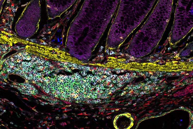
利用Cell DIVE 在单细胞水平上进行超复杂癌症组织分析
能够研究淋巴瘤细胞的异质性如何受到细胞对其微环境反应的影响,尤其是在突变、转录组和蛋白质水平上。蛋白质表达研究提供了有关细胞相互作用性质和蛋白质表达水平的最相关信息。超复合工作流程可用于研究同一癌症组织中的多种蛋白质。
Loading...

Imaging of Anti-Cancer Drug Uptake in Spheroids using DLS
Spheroid 3D cell culture models mimic the physiology and functions of living tissues making them a useful tool to study tumor morphology and screen anti-cancer drugs. The drug AZD2014 is a recognized…
Loading...
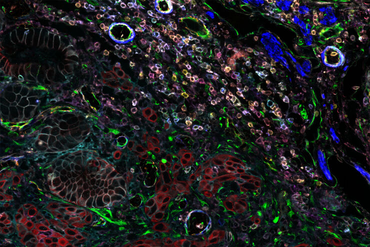
利用多重中频成像设计您的研究课题
多重组织分析是一种功能强大的技术,可对单个固定组织样本中的细胞类型位置和细胞类型相互作用进行比较。在多重分析研究开始之前,研究人员通常会提出以下问题: "我如何知道组织中哪些生物标记物是相关的?另外,随着研究问题的发展,我如何转向其他生物标记物?巧妙的研究设计有助于回答现有的问题,并能继续探索研究开始时并不明显的新联系。
Loading...
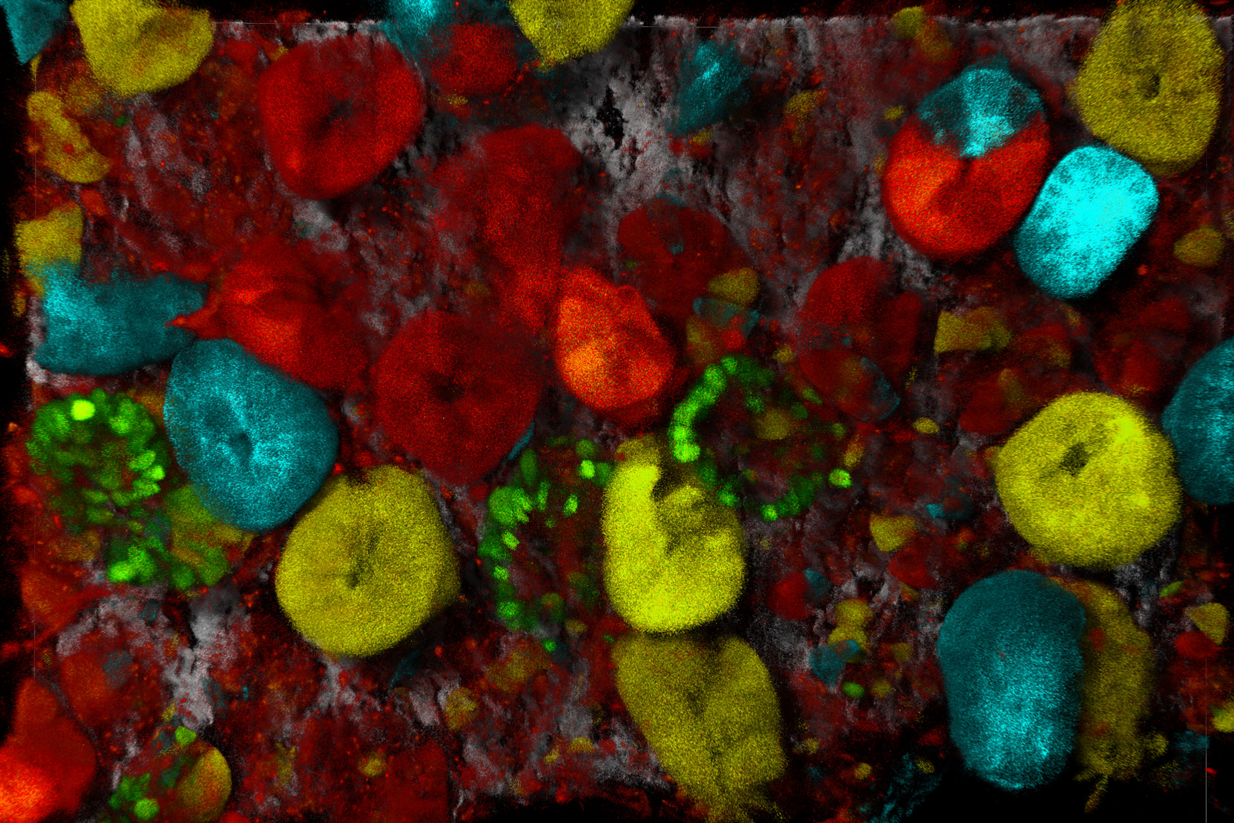
Cell DIVE已验证的抗体将使您对实验结果产生信心
Cell DIVE超多标组织成像分析整体解决方案包括经严格验证的350+抗体资源库,高灵敏度高特异性的应用于Cell DIVE循环染色成像中。抗体验证的方法可以帮助您找到合适的抗体以及最佳的实验条件,快速的让您开展超多标成像分析的实验。抗体库中的每种抗体都经过严格的三步验证过程,(a)评估在FFPE上的表现性能;(b)确定其最佳的染色条件以及是否可用作直标抗体;(c)探究由于Cell…
Loading...
Optimizing THUNDER Platform for High-Content Slide Scanning
With rising demand for full-tissue imaging and the need for FL signal quantitation in diverse biological specimens, the limits on HC imaging technology are tested, while user trainability and…
Loading...
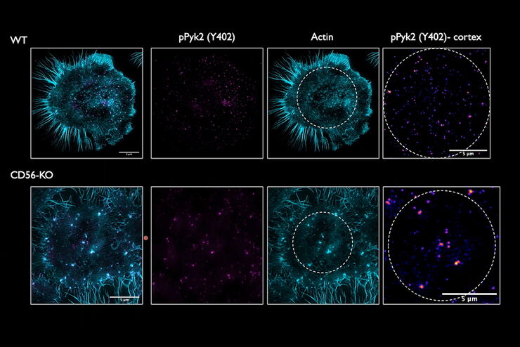
Regulators of Actin Cytoskeletal Regulation and Cell Migration in Human NK Cells
Dr. Mace will describe new advances in our understanding of the regulation of human NK cell actin cytoskeletal remodeling in cell migration and immune synapse formation derived from confocal and…
Loading...
Improvement of Imaging Techniques to Understand Organelle Membrane Cell Dynamics
Understanding cell functions in normal and tumorous tissue is a key factor in advancing research of potential treatment strategies and understanding why some treatments might fail. Single-cell…
Loading...
From Organs to Tissues to Cells: Analyzing 3D Specimens with Widefield Microscopy
Obtaining high-quality data and images from thick 3D samples is challenging using traditional widefield microscopy because of the contribution of out-of-focus light. In this webinar, Falco Krüger…