Gold EM grid with a silicon dioxide (SiO₂) support film, containing vitrified T47D cells intrinsically expressing GFP-H2B (labeling nuclei) and RFP-LifeAct (labeling the actin cytoskeleton) and high-pressure frozen C. elegans germline tissue, fluorescently labeled with GFP-tagged synaptonemal complex protein

EM 시료 준비 워크플로우 & 용도

연구자들은 Leica의 시료 준비 솔루션을 사용함으로써 전자현미경으로 시료를 이미징할 때 고품질, 고정밀, 재현성 있는 결과를 일관되게 얻을 수 있습니다. 당사의 솔루션은 주사전자현미경(SEM), 투과전자현미경(TEM), 극저온전자현미경(Cryo EM)을 지원합니다.

지금 문의하기

당사의 이미징 전문가들은 EM 시료 준비 워크플로우 및 활용 솔루션에 대한 조언을 제공합니다 & 용도

연구자는 SEM 분석을 위해 시료를 어떻게 준비해야 하나요?

SEM(주사 전자 현미경) 시료 준비에는 최적의 이미징을 보장하기 위해 코팅, 건조, 포매와 같은 기법들이 사용됩니다. Leica의 SEM 시료 준비 솔루션을 사용하면 연구자들은 시료의 전도성을 향상시키고, 아티팩트 발생을 최소화하며, 신뢰도 높은 SEM 이미징을 가능하게 하는 안정적이고 고품질의 표면을 확보할 수 있습니다.

TEM 시료 준비에 가장 적합한 방법은 무엇인가요?

TEM(투과전자현미경) 시료 준비에서는 세부 구조를 고해상도로 관찰하기 위해 초박편이면서 손상이 없는 절편 제작, EM 그리드의 스퍼터코팅, 그리고 대비 향상을 위한 염색 과정이 필요합니다. Leica 장비의 정밀성과 신뢰성을 통해 연구자들은 고해상도 TEM 이미지를 얻을 수 있습니다.

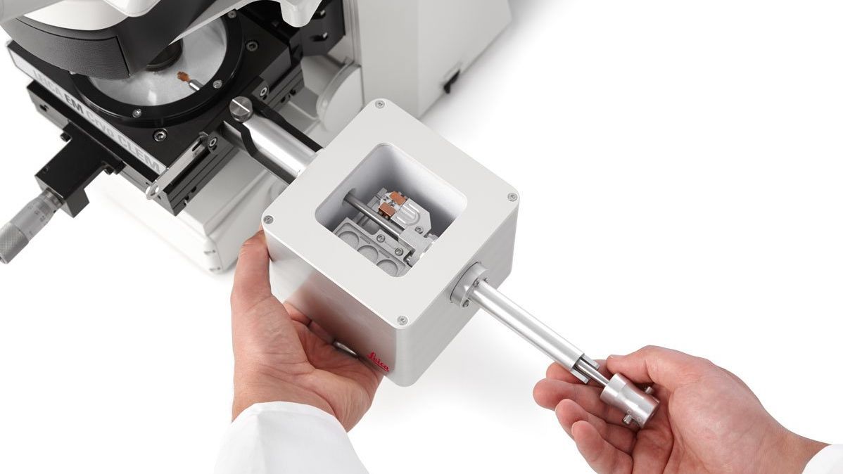
Cryo-EM 시료 준비에서 무엇이 중요한가요?

Cryo-EM에서는 시료 구조를 자연 상태로 보존하기 위해 초고속 냉동을 통한 비정질화(Vitrification)가 핵심 과정입니다.

고급 비정질화(vitrification), 코팅(coating), 초박절편(ultra-thin sectioning), 시료 표면을 저온 상태에서 평탄하게 절삭 처리(Cryo planing), 그리고 사용자 요구에 맞춘 극저온 상태에서 운반 (Cryo transfer) 및 CLEM 워크플로우 등 Leica의 극저온 시료 준비 솔루션은 재현성이 높고 오염이 없는 시료 준비를 가능하게 합니다. 시료의 고유 구조를 유지하고, 정밀한 타겟팅을 제공하며, 고해상도 Cryo EM 이미징을 가능하게 합니다.

EM 시료 준비를위해 Leica의 장비와 솔루션을 선택하는 이유는 무엇인가요?


SEM 시료 준비

Leica Microsystems의 SEM 시료 준비 툴에는 고진공 스퍼터 코터와 임계점 건조기가 포함됩니다. 이러한 솔루션은 고품질의 표면 이미징을 보장합니다. 또한 Leica 장비는 SEM 연구에 일관되고 신뢰할 수 있는 결과를 제공합니다.


TEM 시료 준비

연구자들은 정밀성과 신뢰성을 위해 Leica Microsystems의 TEM 시료 준비 솔루션을 신뢰할 수 있습니다. 초박절편기(ultramicrotome)와 자동 염색 시스템과 같은 필수 장비들은 초박절편을 만들고 고해상도 이미지를 얻는 데 도움을 줍니다. 스퍼터 코팅은 시료 전도성과 이미징 품질을 향상시킵니다.


포괄적인 Cryo-EM 시료 준비

낙하 동결기(plunge freezer)와 고압 동결기(High Pressure Freezer)를 포함하고 있는 Leica Microsystems의 극저온 시료 준비 장비는 빠른 동결과 비정질화(vitrification)를 가능하게 합니다. 또한 당사의 극저온 워크플로우를 위한 시료 운반 시스템은 다양한 종류의 시료에 대한 운반 옵션을 제공합니다. 이 옵션은 시료가 오염되는 것을 방지하며, 정확하고 상세한 Cryo-EM 분석을 가능하게 합니다.

관련 문서

EM 시료 준비 워크플로우에 대한 최신 기사 읽기 & 용도

라이카마이크로시스템즈 지식 포털은 현미경에 관한 과학 연구 및 교육 자료를 제공합니다. 초보자와 숙련된 기술자, 과학자가 일상 업무와 실험에 모두 사용할 수 있는 콘텐츠로 구성되어 있습니다.

더 많은 기사
Sectioning resin-embedded sambles

Ultramicrotomy eBook: Targeting, Trimming & Alignment

Ultramicrotomy is evolving rapidly, and today’s microscopes demand high‑quality sections, precise targeting, and reproducible workflows. This eBook brings together expert application notes, automated…
Foraminifera (Ammonia confertitesta) 
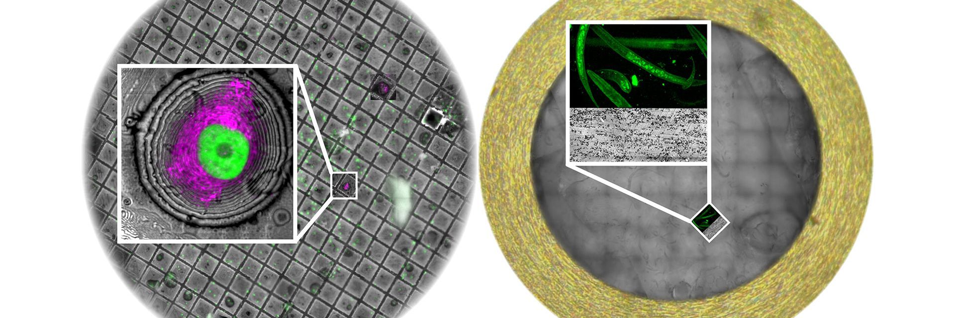
labeled with membrane-permeable calcein, high-pressure frozen in salt water using EM ICE. The sample was cryo-planed and targeted with the M205 on the Cryo-Fluo Enuity, then transferred under cryo conditions to the Cryo-Stellaris for widefield and confocal imaging, revealing details of the staining pattern. Image courtesy: David Evans, University of Southampton.

High-Pressure Freezing for Organoids: Cryo CLEM & FIB Lift Out

Master cryo EM workflow steps for challenging 3D samples: when to choose HPF vs. plunge freezing, reproducible blotting/ice control, contamination aware transfers, Cryo CLEM 3D targeting in organoids,…
Electron microscope (EM) image of a cross section of C. elegans (roundworm). Courtesy of T. Müller-Reichert, MPI-CBG, Dresden, Germany and K. McDonald, University of California, Berkeley, USA.

Brief Introduction to High-Pressure Freezing for Cryo-Fixation

Preparation of biological specimens for electron microscopy (EM) often requires cryo-fixation which does not introduce significant structural alterations of cellular constituents. A common method used…
TEM micrographs of polymer sections. Left: Poly(styrene)-
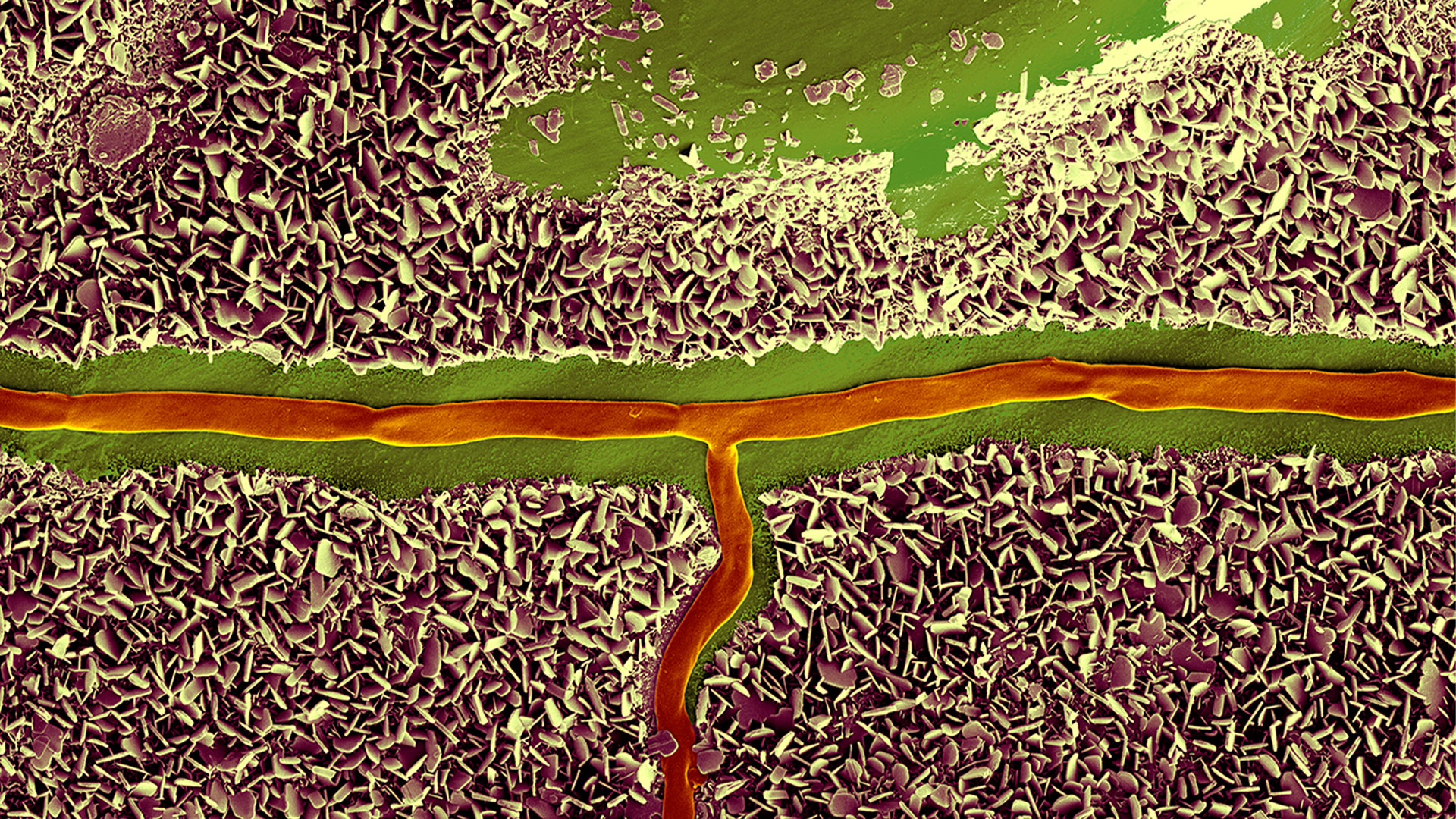
b-poly(isoprene). Right: Poly(styrene)-b-poly(methyl methacrylate).

Ultramicrotome Sectioning of Polymers for TEM Analysis

We demonstrate the capabilities of the UC Enuity ultramicrotome from Leica Microsystems for preparing ultrathin sections of polymer samples under both ambient and cryogenic conditions. By presenting…
Final Segmentation of organelles in Trichomonas species. Magenta – costa, light blue – hydrogenosomes, turquoise – ER, red – vacuoles, yellow – axostyle, green – Golgi apparatus.  Sample courtesy of Isabelle Guerin-Bonne, Low Kay En, Electron Microscopy Unit, Yong Loo Lin School of Medicine, National University of Singapore. Scale bar: 1 µm.

Volume EM and AI Image Analysis

The article outlines a detailed workflow for studying biological tissues in three dimensions using volume-scanning electron microscopy (volume-SEM) combined with AI-assisted image analysis. The focus…

Integrated Serial Sectioning and Cryo-EM Workflows for 3D Biological Imaging

This on-demand webinar explores how integrated tools can support electron microscopy workflows from sample preparation to image analysis. Experts Andreia Pinto, Adrian Boey, and Hoyin Lai present the…
SEM image of the full Li-NMC electrode sample, showing the two porous layers and the metal film at the center of the structure.

Revealing Sodium Battery Degradation via Cryo-EM and CryoFIB

Explore how cryogenic electron microscopy and focused ion beam techniques uncover the intrinsic structure of sodium battery interfaces. This webinar presents a new degradation model based on separator…
Mouse hippocampus brain slice on a grid after HPF using the “Waffle Method”.

The “Waffle Method”: High-Pressure Freeze Complex Samples

This article describes the advantages of a special high pressure freezing method, the so-called “Waffle Method”. Learn how the “Waffle Method” uses EM grids as spacers for high-pressure freezing,…
UC Enuity with Diatome diamond knife.

Mastering Polymer Sectioning with Helmut Gnaegi

When it comes to ultramicrotomy, few names carry the weight of Helmut Gnaegi. As co-founder of Diatome, a global leader in diamond knife technology, Helmut has spent decades refining the art and…
C. elegans embedded in Lowicryl® HM20; pharynx showing red fluorescence (mCherry). The overview shows a front view onto the resin capsule formed by the bottom of a flow-through chamber of the EM AFS2. The capsule was pretrimmed manually. The blockface was trimmed automatically using the AutoTrim function of UC Enuity guided by fluorescence of the worm. Edge length of both squares in relation to the images is 250 µm.

How Fluorescence Guides Sectioning of Resin-embedded EM Samples

Electron microscopes, including transmission electron microscopes (TEM) and scanning electron microscopes (SEM), are widely utilized to gain detailed structural information about biological samples or…
Scroll to top