생명 과학 연구

생명 과학 연구

생명 과학 연구

라이카사의 생명과학사업부는 미세구조의 시각화 및 분석을 위한 혁신 기술 및 기술 전문성을 원하는 scientific community 를 충족시키는 imaging을 지원할 수있습니다. 라이키의 고객을 과학분야의 선두자로 이끌어내는것에 관심이 있습니다.
Mammalian cells expressing H2B-mCherry and alphaTubulin-mEGFP to visualize DNA and microtubules respectively. Left: Widefield imaging using THUNDER Imager. Right: Sample after correlated EM imaging using Leica Microsystems Coral Life workflow. Cells were cryo-immobilized by high-pressure freezing 60 min after the onset of cytokinesis. The intercellular bridge was imaged under TEM after freeze substitution and resin infiltration.

상관관계적 광학 현미경과 전자현미경(CLEM)

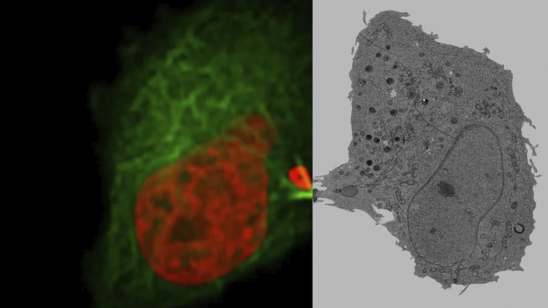
라이카마이크로시스템즈 CLEM 솔루션은 시료 생존성, 품질 확인, 정밀하고 신뢰할 수 있는 3D 타게팅 메커니즘을 보장합니다. 사용자는 적절한 시점에 적합한 시료를 직접 확인하고 높은 분해능의 극저온 공처점 데이터를 얻거나 초미세구조 환경에 형광 정보를 넣기 위해 이 솔루션을 이용할 수 있습니다.

자세히 읽기

Image of C2C12 cells: The cells are stained with lamin B (magenta) which indicates nuclear structure, Hoechst (blue) indicating DNA, and γH2AX (yellow) indicating damage to DNA. Cells were imaged using a THUNDER Imager 3D Live Cell with a 63X/1.4 oil immersion objective.

세포생물학 연구

라이카마이크로시스템즈에서 제공하는 이미징 솔루션은 세포생물학 연구를 최대화하도록 설계되었습니다.

자세히 읽기

세포 배양

실험실 조건에서의 세포 배양은 세포생물학, 암 연구, 발생생물학을 비롯한 모든 종류의 생명 과학 및 제약 연구 분야에 속한 과학자들을 위한 기초입니다. Leica가 실험실에서 동물 세포를 배양하는 데 어떤 도움이 될 수 있는지 알아보십시오.

자세히 읽기

오가노이드와 3D 세포 배양

최근 생명과학 연구에서 가장 흥미로운 발전 중 하나는 오가노이드, 스페로이드 또는 장기 칩 모델과 같은 3D 세포 배양 시스템의 개발입니다. 3D 세포 배양이란 세포가 3차원에서 성장하고 주변 환경과 상호작용할 수 있는 인위적인 환경입니다. 이러한 조건은 체내 상태와 유사합니다.

자세히 읽기

Mammalian cells expressing H2B-mCherry and alphaTubulin-mEGFP to visualize DNA and microtubules respectively. Left: Widefield imaging using THUNDER Imager. Right: Sample after correlated EM imaging using Leica Microsystems Coral Life workflow. Cells were cryo-immobilized by high-pressure freezing 60 min after the onset of cytokinesis. The intercellular bridge was imaged under TEM after freeze substitution and resin infiltration.
Image of C2C12 cells: The cells are stained with lamin B (magenta) which indicates nuclear structure, Hoechst (blue) indicating DNA, and γH2AX (yellow) indicating damage to DNA. Cells were imaged using a THUNDER Imager 3D Live Cell with a 63X/1.4 oil immersion objective.

새로운 Ivesta 3 실체 현미경과 Flexacam 카메라는 Enersight 소프트웨어 기반 포트폴리오를 확장합니다.

QC 커뮤니티를 위한 통합 사용자 인터페이스는 모바일 검사를 가능하게 합니다.

라이카 마이크로시스템즈의 올인원(All-In-One) 디지털 검사 솔루션

Scroll to top