DMi1
도립
광학 현미경
제품소개
홈
Leica Microsystems
DMi1 Inverted Microscope for Cell and Tissue Culture
Just Right – Smart Choice
최신 기사를 읽어 보세요
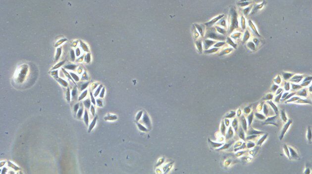
How to do a Proper Cell Culture Quick Check
In order to successfully work with mammalian cell lines, they must be grown under controlled conditions and require their own specific growth medium. In addition, to guarantee consistency their growth…
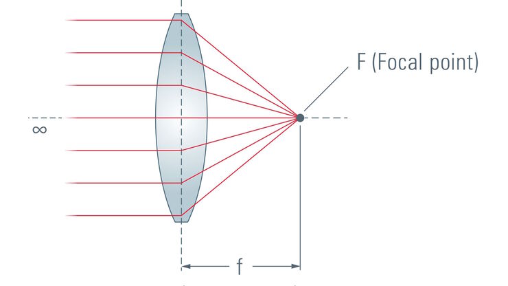
Microscope Resolution: Concepts, Factors and Calculation
This article explains in simple terms microscope resolution concepts, like the Airy disc, Abbe diffraction limit, Rayleigh criterion, and full width half max (FWHM). It also discusses the history.
How to Sanitize a Microscope
Due to the current coronavirus pandemic, there are a lot of questions regarding decontamination methods of microscopes for safe usage. This informative article summarizes general decontamination…
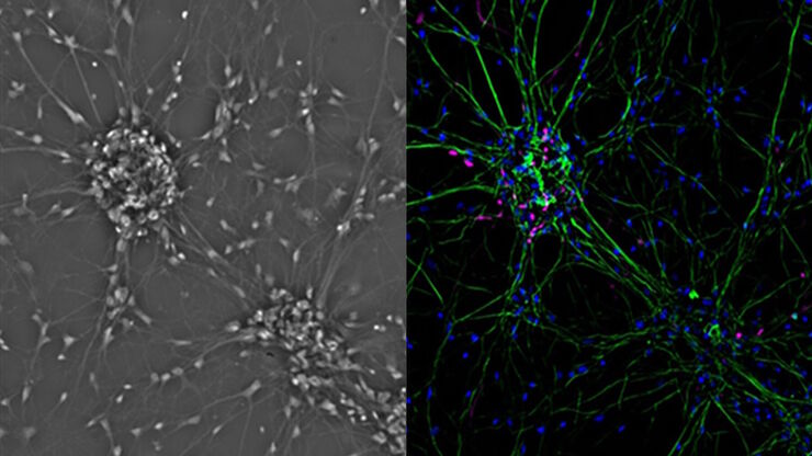
Introduction to Mammalian Cell Culture
Mammalian cell culture is one of the basic pillars of life sciences. Without the ability to grow cells in the lab, the fast progress in disciplines like cell biology, immunology, or cancer research…
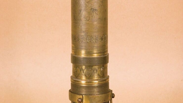
A Brief History of Light Microscopy
The history of microscopy begins in the Middle Ages. As far back as the 11th century, plano-convex lenses made of polished beryl were used in the Arab world as reading stones to magnify manuscripts.…
Optical Microscopes – Some Basics
The optical microscope has been a standard tool in life science as well as material science for more than one and a half centuries now. To use this tool economically and effectively, it helps a lot to…
Fields of Application
세포 배양
실험실 조건에서의 세포 배양은 세포생물학, 암 연구, 발생생물학을 비롯한 모든 종류의 생명 과학 및 제약 연구 분야에 속한 과학자들을 위한 기초입니다. Leica가 실험실에서 동물 세포를 배양하는 데 어떤 도움이 될 수 있는지 알아보십시오.
