is mobile? false
Microscope Products
Microscope Products
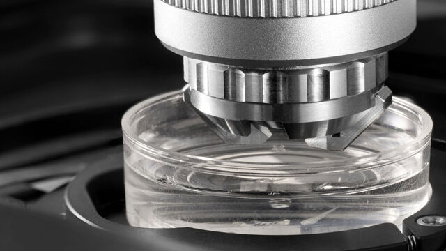
We develop high-tech precision microscopes for the analysis of microstructures with the user, for the user. In our product portfolio you will find microscopes for life science including biotechnology and medicine as well as for research and development of raw materials and industrial quality assurance.
Product Categories Show subnavigation
Microscope Products
High-tech precision microscopes for the analysis of microstructures. Find microscope solutions for life science, medical, R&D and industrial quality assurance.


Our Latest Innovations Show subnavigation


The Award-Winning Inspection Microscope
The Emspira 3, which integrates comparison, measurement, and documentation sharing into a single system, has won a prestigious Red Dot Award.


Upcoming Events Show subnavigation
Take a look at all our upcoming congresses, exhibitions, webinars, and workshops and join us at one of our next events!
Applications Show subnavigation


Leica Science Lab Articles Show subnavigation
Read our latest articles
The knowledge portal of Leica Microsystems offers scientific research and teaching material on the subjects of microscopy. The content is designed to support beginners, experienced practitioners and scientists alike in their everyday work and experiments.
How Fluorescence Guides Sectioning of Resin-embedded EM Samples
Electron microscopes, including transmission electron microscopes (TEM) and scanning electron microscopes (SEM), are widely utilized to gain detailed structural information about biological samples or non-living materials. Ultramicrotomy is the preferred technique for producing ultrathin sections, less than 100 nm thick for TEM/SEM analysis. During sample preparation small sample pieces are embedded in epoxy or acrylic resin, excess resin is trimmed away, and the specimen is sliced into ultrathin sections (50 nm - 100 nm) using a glass or diamond knife.
Coherent Raman Scattering Microscopy Publication List
CRS (Coherent Raman Scattering) microscopy is an umbrella term for label-free methods that image biological structures by exploiting the characteristic, intrinsic vibrational contrast of their molecules. The two most important CRS techniques are Coherent Anti-Stokes Raman Scattering (CARS) and Stimulated Raman Scattering (SRS). The biochemical image contrast of CRS is in many ways complementary to the molecular contrast obtained in fluorescence microscopy. A second crucial advantage of these methods is that they preserve the specimen/sample in a near pristine state. This reference list presents current and basic papers on CRS microscopy.
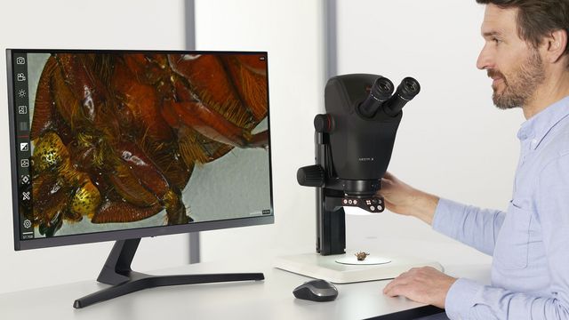
Selecting the Right Dissecting Microscope
Learn how you can enhance dissection for life-science research and education with a microscope that ensures ergonomic comfort, high-quality optics, and easy access to the specimen.

A Guide to Polarized Light Microscopy
Polarized light microscopy (POL) enhances contrast in birefringent materials and is used in geology, biology, and materials science to study minerals, crystals, fibers, and plant cell walls.
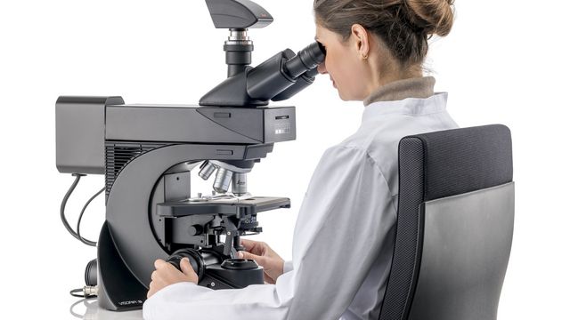
Factors to Consider when Selecting Clinical Microscopes
What matters if you would like to purchase a clinical microscope? Learn how to arrive at the best buying decision from our Science Lab Article.
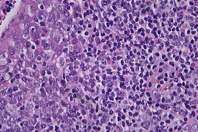
Clinical Microscopy: Considerations on Camera Selection
The need for images in pathology laboratories has significantly increased over the past few years, be it in histopathology, cytology, hematology, clinical microbiology, or other applications. They serve many purposes on top of the documentation of the diagnosis. Yet, the view through the eyepieces and the image are different in nature, the one is an optical image, the other a digital image. Looking at a few aspects of this process that are related cameras will help you make sure you can obtain the images with all the detail and color fidelity you need.
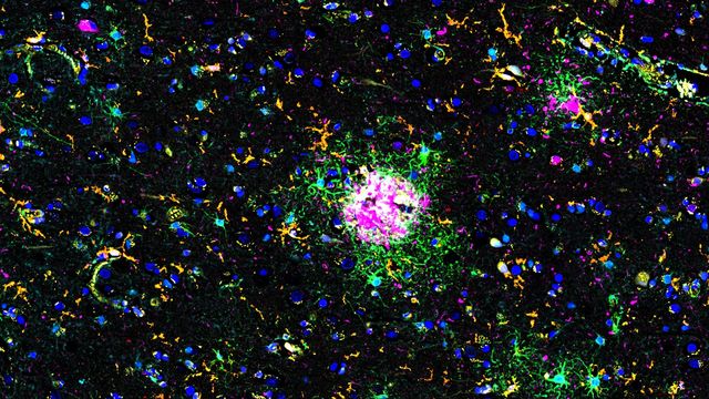
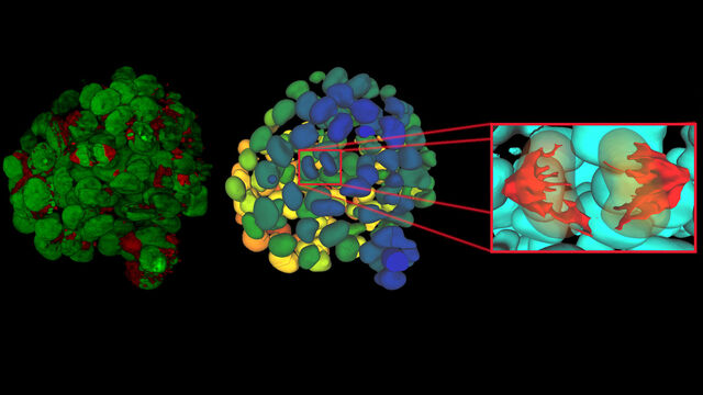
Explore Alzheimer's Spatial Proteome with Big Data
Alzheimer's disease, a genetic and sporadic neurodegenerative condition, leads to cognitive decline in mid to late life, marked by β-amyloid plaques and tau tangles. With limited treatment options, new investigative strategies are crucial. The Cell DIVE multiplexed imaging solution allows examination of Alzheimer's brain tissue, potentially uncovering new research avenues. Here, we showcase the Cell DIVE image viewer, enabling users to access the full Alzheimer's multiplexed dataset directly in their browser.
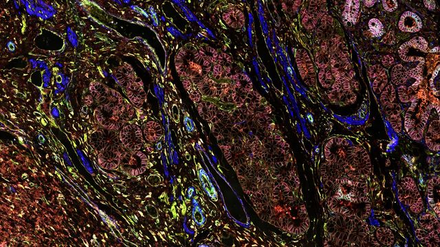
Dive into Pancreatic Cancer Research with Big Data
Pancreatic cancer, with a mortality rate near 40%, is challenging to treat due to its proximity to major organs. This story explores the complex biology of pancreatic ductal adenocarcinoma (PDAC), examining molecular and spatial determinants of tumor aggression in metabolism, apoptosis, and immunity. Access the full Cell DIVE dataset in your browser to delve deeper into these findings.
