Electron Microscope Sample Preparation

Electron Microscope Sample Preparation

Electron Microscope Sample Preparation

Excellent sample preparation is the prerequisite for first-class electron microscopy. Be prepared – for great results in EM Sample Preparation! Perfect preparation makes the difference between trying and achieving, between failure and success, between results and excellent results. So be prepared for great results with Leica Microsystems!

What are you interested in?

EM GP2

Automatic Plunge Freezer

STELLARIS Cryo

STELLARIS Cryo is a confocal light microscope system that helps you to target your area of interest for cryo-electron tomography (CryoET)

UC Enuity

Next generation ultramicrotome that saves you valuable time and resources through its automated setup functions, providing state-of-the-art sectioning quality.

EM TP

With the automated EM TP Tissue Processor, ultrastructures of your tissue samples can be precisely prepared every time.

EM ACE200

Low vacuum sputter and carbon thread coater

EM ACE600

High vacuum sputter, carbon thread and e-beam coater

EM ACE900

High-End EM Sample Preparation Freeze Fracture System

EM CTD

Fast, Reliable Cryo Tool Drying for Efficient Cryo EM workflow

EM CPD300

Critical Point Dryer

EM AFS2

Freeze Substitution and Low Temperature Embedding System for Light and Electron Microscopy

EM RAPID

Trim your biological and industrials samples fast and accurately with the EM RAPID high-speed milling system.

EM AC20

Automatic contrasting instrument for ultrathin sections

EM TXP

Mechanical cross-sectioning device that precisely downsizes samples for visual inspection. Optimize sample pre-preparation to minimize ion beam milling time and ensure creation of mirror-like surfaces...

Read our latest articles about Electron Microscope Sample Preparation

The knowledge portal of Leica Microsystems offers scientific research and teaching material on the subjects of microscopy. The content is designed to support beginners, experienced practitioners and scientists alike in their everyday work and experiments.

More Articles

How to Save Time and Samples by Automated Ultramicrotomy

This article describes how 3D micro-CT data of a resin-embedded electron microscopy sample can be used to trim the specimen down to a defined target plane prior to sectioning. The interactive and automated approach described using Leica Microsystems’ UC Enuity saves time, reduces sample loss and training time for unexperienced users.

From Bench to Beam: A Complete Correlative Cryo Light Microscopy Workflow

In the webinar entitled "A Multimodal Vitreous Crusade, a Cryo Correlative Workflow from Bench to Beam" a team of experts discusses the exciting world of correlative workflows for structural biology that empower researchers to study fine details of biological structures. Watch and explore the latest developments, instruments, and techniques in cryo workflows for correlative light electron microscopy (cryo-CLEM).
Feb 06, 2025
Webinar
CLEM

A Guide to Cryo-Electron Tomography

Cryo-electron tomography (CryoET) is used to resolve biomolecules within their cellular environment down to an unprecedented resolution below one nanometer.

How to Automatically Obtain Fluorescent Cells of Interest in a Block-face

Block-face created by automatic trimming under fluorescence. Mammalian cells of interest, stained with CellTrackerTM Green are visualized within the block-face using the UC Enuity equipped with the stereo microscope M205 FA. In the background a carbon finder grid in black is visible.

Improve Your Ultramicrotomy Workflow with Automated Sectioning

Discover advanced digital ultramicrotomy tools for fast and accurate automated sectioning. Learn about autoalignment, and efficient sample trimming leveraging 3D µCT data. See application examples that advance EM sample preparation workflows. Watch now to enhance your lab's precision and efficiency.

Workflow Solutions for Sample Preparation Methods for Material Science

This brochure presents and explains appropriate workflow solutions for the most frequently required sample preparation methods for material science samples.

Automatic Alignment of Sample and Knife for High Sectioning Quality

Automatic alignment of sample and knife on the ultramicrotome UC Enuity, enabling even untrained users to create ultrathin sections with reduced risk of losing precious sections.

High Quality Sectioning in Ultramicrotomy

Discover the significance of achieving high-quality uniform sections with ultramicrotomy for precise imaging in electron microscopy.

Rapid Semiconductor Inspection with Microscope Contrast Methods

Semiconductor inspection during the production of patterned wafers and ICs (integrated circuits) is important for identifying and minimizing defects. To increase the efficiency of quality control in the early stages of production and to ensure reliable IC chip performance, microscopy solutions should combine different contrast methods that provide complete and accurate information about different defects. Our free guide details microscopy techniques & optimizing quality control. Get your free copy today!

Five Inverted-Microscope Advantages for Industrial Applications

With inverted microscopes, you look at samples from below since their optics are placed under the sample, with upright microscopes you look at samples from above. Traditionally, inverted microscopes are used for life science research, because gravity makes samples sink to the bottom of a holder with aqueous solution and you don’t see a lot from above.

Structural and Chemical Analysis of IC-Chip Cross Sections

This article shows how electronic IC-chip cross sections can be efficiently and reliably prepared and then analyzed, both visually and chemically at the microscale, with the EM TXP and DM6 M LIBS solutions.

High-Quality EBSD Sample Preparation

This article describes a method for EBSD sample preparation of challenging materials. The high-quality samples required for electron backscatter diffraction are prepared with broad ion-beam milling.

How Marine Microorganism Analysis can be Improved with High-pressure Freezing

In this application example we showcase the use of EM-Sample preparation with high pressure freezing, freeze substiturion and ultramicrotomy for marine biology focusing on ultrastructural analysis of dinoflagellates in particular. The mobility the EM ICE unit allowed high pressure freezing directly at the marine station after sample collection.
Aug 07, 2023
Tutorial
CLEM

How to Prepare and Analyse Battery Samples with Electron Microscopy

This workshop covers the sample preparation process for lithium and novel battery sample analysis, as well as other semiconductor samples requiring high-resolution cross-section imaging.

New Imaging Tools for Cryo-Light Microscopy

New cryo-light microscopy techniques like LIGHTNING and TauSense fluorescence lifetime-based tools reveal structures for cryo-electron microscopy.

How to Target Fluorescent Structures in 3D for Cryo-FIB Milling

This article describes the major steps of the cryo-electron tomography workflow including super-resolution cryo-confocal microscopy. We describe how subcellular structures can be precisely located in 3D under cryo-fluorescence and how their coordinates are provided for subsequent cryo--FIB milling and cryo-transmission electron microscopy.
Leica EM VCT500 & Leica EM ACE600

Sample Preparation

In the field of electron microscopy, perfect sample preparation is a prerequisite and crucial step. Leica Microsystems offers a comprehensive product portfolio for preparation of biological, medical and industrial samples. Concentrating on workflow solutions we provide a product range that is perfectly aligned to all your needs for precise sample preparation in TEMSEM, and AFM investigations. Each Leica solution consists of several instruments that are perfectly geared to one another to form a seamless workflow for your sample.
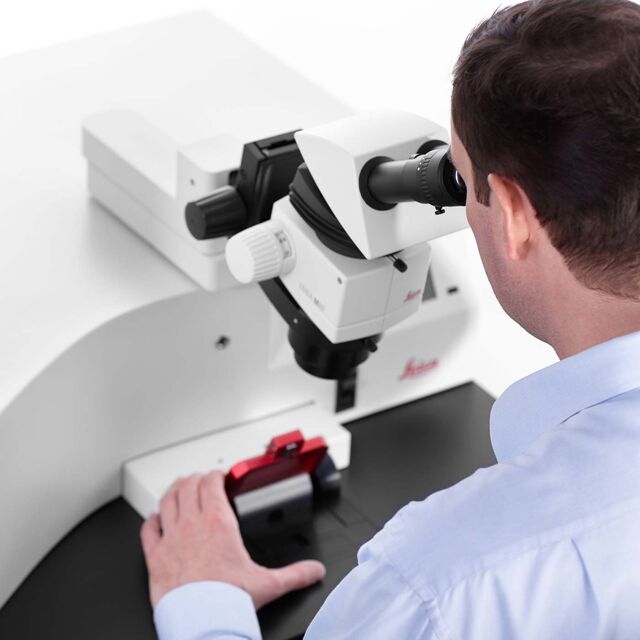
Volume reconstruction of a book scorpion (outer view and inner view to visualize the musculature).

Critical Point Dryer

Fully automated and controlled process!

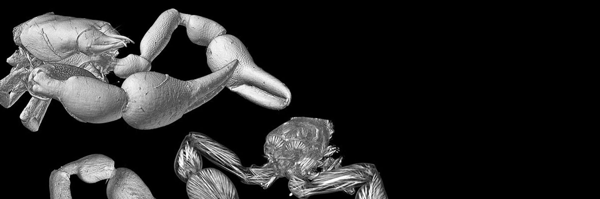
The procedure of critical point drying is an efficient method for drying delicate samples for SEM applications. It preserves the surface structure of a specimen which could otherwise be damaged due to surface tension when changing from the liquid to gaseous state. The Leica EM CPD300 critical point dryerdries biological specimens such as pollen, tissue, plants, insects, as well as industrial samples, like Micro Electro Mechanical Systems (MEMS) or Micro fluids and gels in a fully automated and controlled process. You can rely on the same high sample quality from every run.

Leica EM VCT500

Sample Transfer System

Seamless workflows!

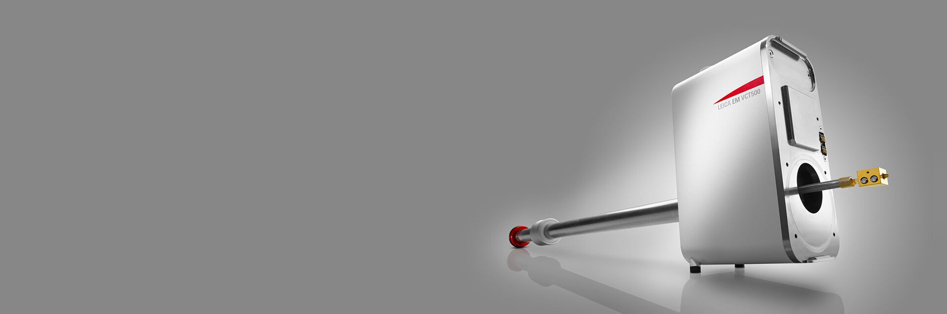
When transferring specimens to the chamber of an analysis system it is essential to protect the samples from contamination. Concentrating on workflow solutions, Leica Microsystems has developed a sophisticated vacuum transfer system which provides transfer options for all kind of samples. Users can thus rely on a system that works seamlessly without interruption: The transport systems and Leica EM VCT500 serves as a shuttle from one step to the next, always preserving cryo and / or vacuum conditions.
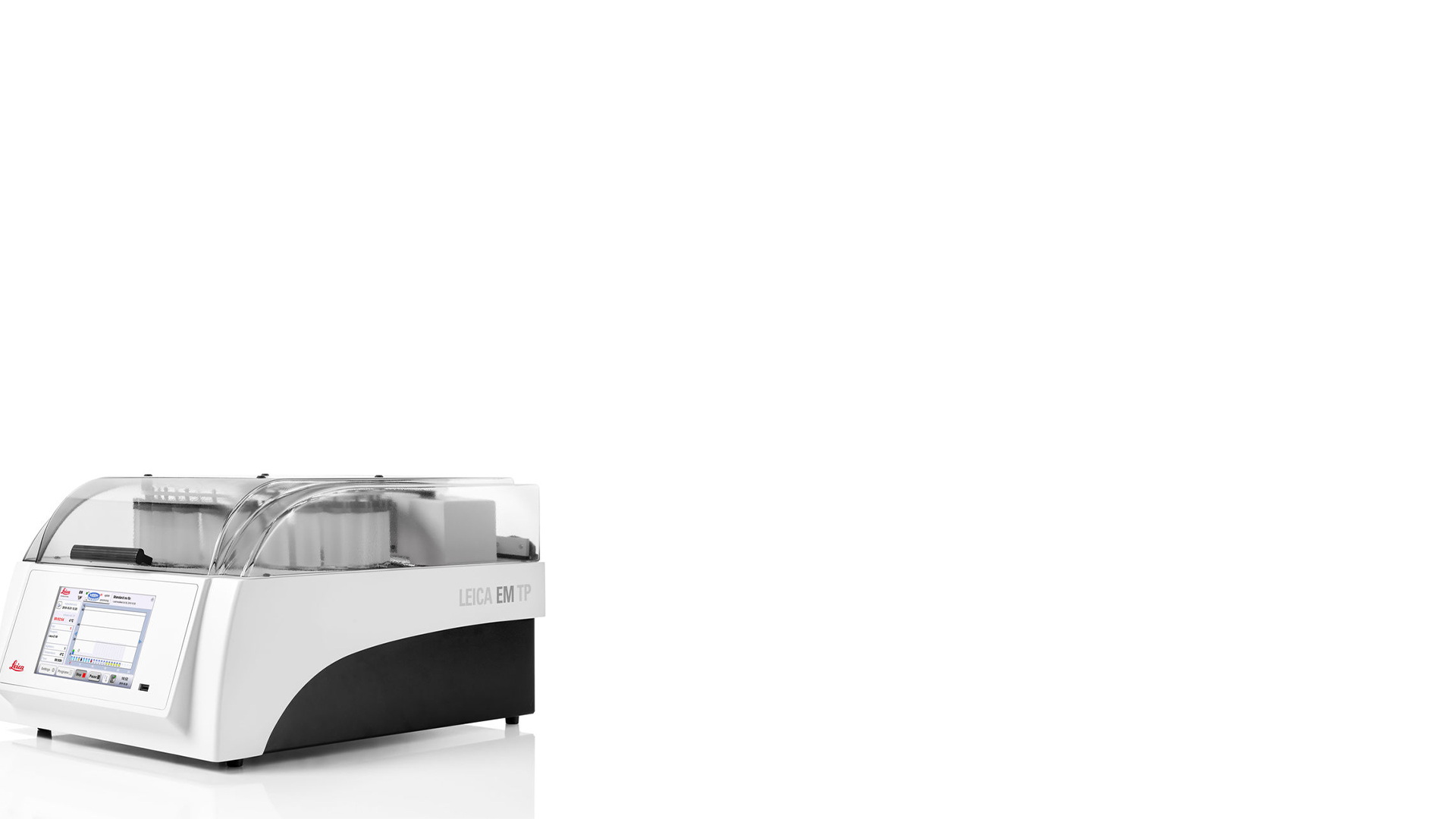
Automated Tissue Processor EM TP

Tissue Processor

Only correctly prepared tissues provide useful microscopic information. Leica Microsystems’ tissue processors reduce the risk of an inappropriate handling by automation as well as increasing efficiency, making sure you can reproduce the process at any time.

Reduce Processing Time

With minimal user interaction, the Leica EM TP processes and embeds into resin.

Increase User Safety

Automatic processing of tissue minimizes contact with hazardous reagents, provides reproducible results, increases time savings and improves user safety in the laboratory. The Leica EM TP tissue processor, designed for EM and LM resin processing, features an exhaust system that supports safer use of toxic substances.

Trimming Systems

For biological, industrial or pharmaceutical applications!

High Preparing embedded samples for ultramicrotomy is a delicate task. As the shape of the block face and the straightness of the edges of the trimmed sample have a profound effect on the sectioning characteristics, parallel edges top and bottom are a must.

Leica Microsystems’ trimming devices for biological, industrial or pharmaceutical applications make the first step of sample preparation safe, accurate, fast and reliable. Users who prepare TEM specimens for ultrathin sections obtain surfaces of the highest sectioning quality. In pharmaceutical research pills can be milled with maximum safety and precision.

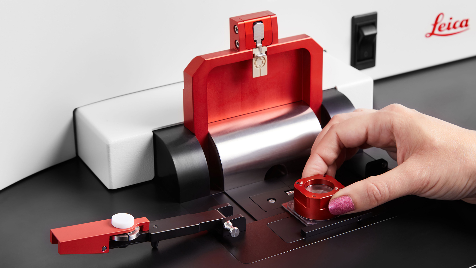
Pharmaceutical Research

In the development of new medications, the distribution of active ingredients within a tablet plays an important role in their effectiveness. Prepare tablets for quantitative NIR (near-infrared) spectroscopy quickly and with maximum precision using the Leica EM RAPID milling system. High-performance tungsten carbide or diamond milling tools decapsulate and mill without smearing effects from the outer capsule layer.

Trimming for SEM, TEM and LM

Conventional trimming of embedded samples involves the use of razor blades and a great deal of skill by the ultramicrotomist. The Leica EM RAPID specimen trimming device helps you to produce a perfect pyramid and cutting face of both biological and industrial samples safely, rapidly and accurately within less than 60 seconds.

The EM ICE Nano loading area

Cryo Preparation Systems

Visualize highly dynamic processes!

High pressure freezing is often the preferred method for preserving aqueous samples in their close-to-native state, as it captures the intricate changes in fine structure or cellular dynamics. Leica Microsystems combines high pressure freezing with light stimulation or electrical stimulation: It enables you to visualize highly dynamic processes or the structural changes of samples at a nanometer resolution and with millisecond precision. This allows researchers in life sciences and industry to get answers to questions for which they were unable to design experiments before.

Fully Integrated Light Stimulation

The Leica EM ICE high pressure freezer is the only instrument capable of synchronizing freezing and stimulation with a millisecond precision. Apply light stimulation to any light sensitive compound such as lotions, cosmetics, or food, and photo-activated samples such as proteins, or various biological samples.

Fluid or Thin Sample Preparation

To prepare vitrified thin samples for cryo-TEM, including biological suspensions and industrial emulsions in both aqueous and inorganic solvents, the bare grid technique is used. The Leica EM GP2 plunge freezes fluid or extremely thin samples spread on an electron microscopy grid into liquid ethane after removing excess fluid by automatic blotting.

Freeze Substitution at Low Temperature

Freeze Substitution is a common follow-on procedure to high pressure freezing and other cryo fixation methods. It is the process of dehydration of the frozen specimen by an organic solvent at low temperature in the presence of a secondary fixative. The Leica EM AFS2 freeze substitution and low temperature embedding system features a remote control that makes it easy to access your system at any time from any location, and be in control of your experiment and laboratory.

Electron Microscopy Sample Preparation Products 15

EM GP2

Automatic Plunge Freezer
STELLARIS Cryo Confocal Microscope

STELLARIS Cryo

STELLARIS Cryo is a confocal light microscope system that helps you to target your area of interest for cryo-electron tomography (CryoET)
Ultramicrotome UC Enuity

UC Enuity

Next generation ultramicrotome that saves you valuable time and resources through its automated setup functions, providing state-of-the-art sectioning quality.

Education

Educational microscope solutions from Leica Microsystems for school and university feature optical performance and student-friendly construction.

Read More

Life Science Research

Leica Microsystems’ life science research microscopes support the imaging needs of the scientific community with advanced innovation and technical expertise for the visualization, measurement and analysis of microstructures.

Read More

Materials & Earth Science

Physicals science research microscopes from Leica Microsystems offer a broad range of optics, imaging systems, software, and ergonomic accessories to design a system for your application.

Read More

Industrial Applications

Microscopes for Industrial Applications from Leica Microsystems help you minimize downtime through a broad range of solutions and expert local support.

Read More

Education

Educational microscope solutions from Leica Microsystems for school and university feature optical performance and student-friendly construction.

Read More

Life Science Research

Leica Microsystems’ life science research microscopes support the imaging needs of the scientific community with advanced innovation and technical expertise for the visualization, measurement and analysis of microstructures.

Read More

Interested to know more?
Talk to our experts.

Do you prefer personal consulting? Show local contacts

Scroll to top