Modulation contrast
The modulation contrast is a contrast enhancing method in wide-field microscopy that converts optical gradients or slopes in unstained specimen and live cells into varying light intensities. It was invented by Robert Hoffman in 1975 and employs a slit plate to enable oblique illumination and a modulator for light intensity modulation. Due to the three dimensional appearance of the specimen, main applications of modulation contrast in medicine and life sciences are micromanipulation techniques like ICSI (intracytoplasmic sperm injection) in IVF and cloning and pronucleus injection in transgenics.
Special components in the light path
At least two special components in the light path are necessary for modulation contrast illumination. To enable oblique illumination a slit plate is placed in the front focal plane of the condenser. Secondly, a modulator is placed in the back focal plane of the objective. The modulator contains three designated areas with different optical densities: one transparent area, one with 15 % light permission and one with 1 % light permission. Light passing the modulator is by definition called modulated.
In addition to the above mentioned components a second slit containing a polarizer can often be found in the slip plate. In combination with another polarizer located on top of the condenser of the microscope, this polarizer can be used to control the effective slit opening (by opening the 90° (dark) position) and enable relief effects. Note that the specimen is not located between the polarizers which allows the employment of birefringent plastic or glass vessels. However, the polarizers are not a prerequisite for modulation contrast.


Modulation contrast converts phase gradients to brightness gradients
Many components of cells cause changes in the phase of transmitted light by their innate properties. These properties might be the slope and the steepness of a specific structure, a structure that modifies the rate of change in refractive index or simply changes in the thickness of a given structure. As these properties (e.g. cell thickness) often change gradually over a certain area (instead of changing instantly), the change of the phase of light over this area also occurs gradually resulting in a phase gradient. Phase gradients will diffract the incident light which causes a change in the angle of deflection of the light.
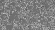
Due to the oblique illumination that is employed in modulation contrast, the change in the angle of deflection might greatly be affected and the light will pass the modulator in a different area compared to non-deflected light. In the resulting images areas of the cell(s) that are more or less homogenous in turns of slope, steepness, thickness etc. will be displayed in grey. The same is true for the background of the image, as light passing these homogenous areas does not experience a phase shift due to phase gradients in the specimen and will hardly be deflected.
In a modulation contrast system non-deflected light passes the area in the modulator with 15 % light permission, resulting in grey color for homogenous areas of the cell(s) and the background. When light passes areas of the specimen that cause a phase shift (e.g. changes in the slope of a cell membrane), the light is deflected either to the dark (1 % permission) or the transparent (100 % permission) area of the modulator. As the specimen is obliquely illuminated, the image will appear darker from one side of the gradient (where the light is deflected to area with 1 % permission) and brighter from the other side of the gradient (where the light is deflected to the area with 100 % permission).

IMC offers more freedom
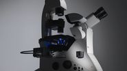
In the case of Integrated Modulation Contrast (IMC) the modulator is integrated outside the objective in a focal plane conjugate to the condenser (intermediate pupil) in the beam path (Figure 4). The modulators are mounted on sliders which are inserted into the interpupillary interface on the side of the stand (Figure 5). They are therefore freely accessible and allow the user to easily adjust the contrast as required. Beyond this, it is also possible to use modulators with different transmissions. The slider also features a brightfield position for unmodulated images. Microscopes with IMC have been set up to ensure that the orientation of the contrast (shadow direction of the 3D impression) is identical for all objectives. The spatial impression of the specimen thus remains unchanged when changing objectives or magnifications, so there is no need to adjust the slit diaphragm.


A fundamental consideration when uncoupling the modulator was that the rear focal plane of the objective can be anywhere within the entire range of the 45 mm parfocality distance. A modulator outside of the objective must therefore be movable by at least that distance. Thanks to the design of the Leica objectives, which is defined by fixed focal planes, deployment in inverted microscopes and fixed magnification steps from 5x to 63x, it was possible to reduce the number of modulation planes to two pupil positions (Figure 6).
The beam path to the image of the pupil, the so-called intermediate pupil, is designed as a 1x telescope system. The infinite beam path behind the objective is reproduced in the intermediate pupil. As a result, plane-parallel optical modulators of any thickness can also be inserted into the beam path at the intermediate pupil, tilted or untilted, without impairing the position or quality of the image. Stray and false light are minimized by the optimal monochromatic and chromatic correction of the telescope system as well as the extremely low reflection properties of the vapour-deposited grey and dark metal coatings of the IMC modulator.

Individually variable parameters
The IMC offers plenty of scope for individual adjustment of the modulation contrast. The modulator is adjustable vertically to provide a homogeneous image at all times. Varying stage or buffer solution levels and different positions of the cells within the specimen dish can thus be compensated. Moving the modulator sideways alters the resolution: The further the modulator and the slit diaphragm are moved outwards, the higher the resolution (max. resolution Res = λ\2NA).
Two modulator parameters affect the strength of the contrast: the transmission and the width of the grey area. Reducing transmission increases contrast. Transmission of 25 %, for example, results in smooth contrast. With a value of around 10 %, the contrast becomes significantly crisper. Varying the modulator width is similarly effective (Figure 7).

Narrowing the grey area results in crisper contrast, while wide modulators soften the overall image impression. The slit diaphragm of the IMC features two openings. The first slit is imaged on the grey area of the modulator, producing the oblique illumination for the modulation contrast. The second slit is covered by a polarization foil (Figure 7). A rotatable polarizer positioned in front of the diaphragm can increase or decrease the 3D effect of the modulation contrast. The polarizer does not affect image quality when working with plastic dishes, as the light is polarized only on the illumination side and no analyzer is used on the objective side. The sizes of the slit diaphragms are matched to the most commonly used condensers.
IMC is therefore not only suitable for routine examinations of biological material – it is also an excellent tool for demanding applications such as micromanipulation, microinjection or microdissection.
Advantages of IMC
Numerous advantages make integrated modulation contrast a widely used method in wide field microscopy. Integrated modulation contrast allows the usage of objectives with magnification factors up to 63X. No special objectives are needed and objectives with high correction (e.g. planachromatic and apochromatic objectives) can be employed for modulation contrast. This results in excellent resolution of details with good contrast and visibility
Another advantage of modulation contrast microscopy is that, unlike in differential interference contrast microscopy, plastic vessels and dishes can be used, as the polarizers in the light path are located before the sample. Furthermore no Wollaston prisms are needed. This results in moderate costs for a microscope equipped with modulation contrast.
Reference
Hoffman, R.: The Modulation Contrast Microscope: Principles and Performance, Journal of Microscopy, Vol 110, pt 3, August 1977, pp. 205–222
, tubulin with Cy5 (red), and nuclei with DAPI (blue). Image courtesy of Dr. Günter Giese, Max Planck Institute for Medical Research, Heidelberg, Germany.")
, Tropomyosin (cardiomyocytes, red) and GFP (primordial cardiac layer, green).")