STELLARIS CRS
공초점 레이저 현미경
제품소개
홈
Leica Microsystems
STELLARIS CRS CRS 현미경
무염색 화학 현미경 알아보기
최신 기사를 읽어 보세요
 dataset, showing the biochemically distinct structures of a fresh, untreated apple slice.")
How to Prepare Samples for Stimulated Raman Scattering (SRS) imaging
Find here guidelines for how to prepare samples for stimulated Raman scattering (SRS), acquire images, analyze data, and develop suitable workflows. SRS spectroscopic imaging is also known as SRS…
, unsaturated lipids (magenta, 3050 cm-1), collagen (SHG, cyan). Sample courtesy of R. Rudolf, J Klicks, Hochschule Mannheim")
The Potential of Coherent Raman Scattering Microscopy at a Glance
Coherent Raman scattering microscopy (CRS) is a powerful approach for label-free, chemically specific imaging. It is based on the characteristic intrinsic vibrational contrast of molecules in the…

Formulated Product Characterization with SRS Microscopy
Creams, pastes, gels, emulsions, and tablets are ubiquitous across a wide range of manufacturing sectors from pharmaceuticals and consumer health products to agrochemicals and paint. To improve…

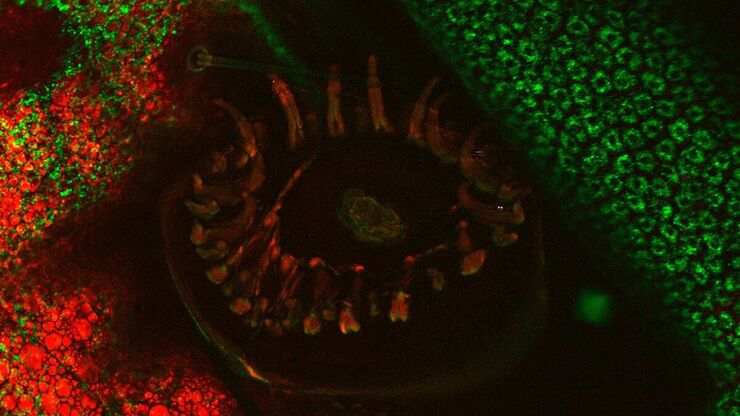
연구 분야의 모델 유기체
모델 유기체는 연구자들이 특정한 생물학적 과정을 연구하기 위해 사용하는 종입니다. 이들은 인간과 유사한 유전적 특성을 가지고 있으며, 유전학, 발달생물학, 신경과학 같은 연구 분야에서 일반적으로 사용됩니다. 유기체 모델은 일반적으로 실험실 환경에서 쉬운 유지와 번식, 짧은 세대 주기 또는 특정 형질이나 질병을 연구하기 위한 돌연변이 생성 능력 때문에…

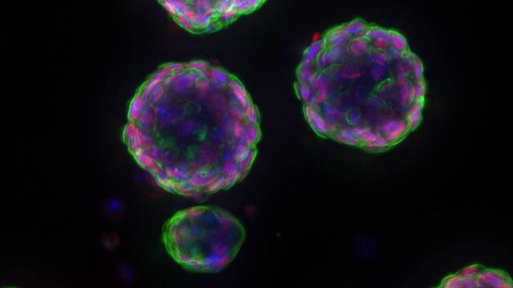
암 연구
암은 성장 통제에 결함이 있는 세포에 의해 발생하는 복잡하고 이질적인 질병입니다. 하나 또는 한 그룹의 세포에서 일어나는 유전적 또는 후생적 변화가 정상적인 기능을 방해하고, 자율적이고 통제되지 않는 세포 성장과 증식을 초래합니다
Stimulated Raman Scattering Microscopy Probes Neurodegenerative Disease
Despite decades of research, the molecular mechanisms underlying some of the most severe neurodegenerative diseases, such as Alzheimer’s or Parkinson’s, remain poorly understood. The progression of…

CARS Microscopy: Imaging Characteristic Vibrational Contrast of Molecules
Coherent anti-Stokes Raman scattering (CARS) microscopy is a technique that generates images based on the vibrational signatures of molecules. This imaging methods does not require labeling, yet…
An Introduction to CARS Microscopy
CARS overcomes the drawbacks of conventional staining methods by the intrinsic characteristics of the method. CARS does not require labeling because it is highly specific to molecular compounds which…
Biopharma
For biopharma, Leica solutions help accelerate drug discovery, enhance cellular analysis, and support data integrity that meets regulations.
