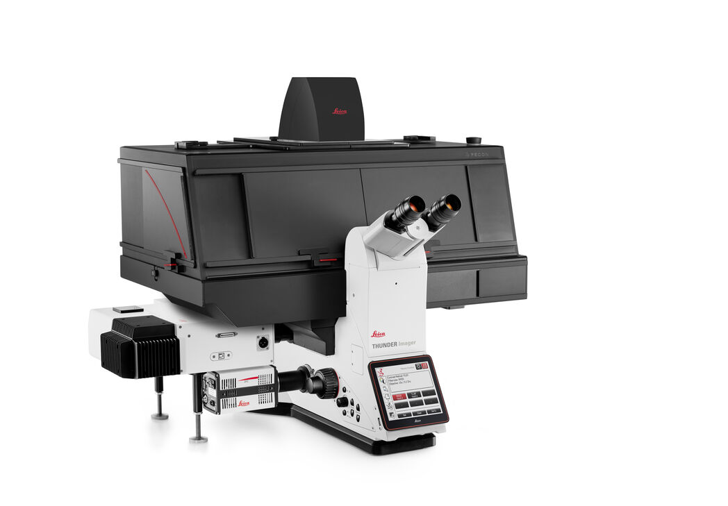
DMi8 S 플랫폼
도립
광학 현미경
제품소개
홈
Leica Microsystems
DMi8 S 플랫폼 도립 현미경 솔루션
미싱 링크 찾기
최신 기사를 읽어 보세요
Studying Virus Replication with Fluorescence Microscopy
The results from research on SARS-CoV-2 virus replication kinetics, adaption capabilities, and cytopathology in Vero E6 cells, done with the help of fluorescence microscopy, are described in this…
Epi-Illumination Fluorescence and Reflection-Contrast Microscopy
This article discusses the development of epi-illumination and reflection contrast for fluorescence microscopy concerning life-science applications. Much was done by the Ploem research group…
Introduction to Fluorescent Proteins
Overview of fluorescent proteins (FPs) from, red (RFP) to green (GFP) and blue (BFP), with a table showing their relevant spectral characteristics.
Differential Interference Contrast (DIC) Microscopy
This article demonstrates how differential interference contrast (DIC) can be actually better than brightfield illumination when using microscopy to image unstained biological specimens.
Phase Contrast and Microscopy
This article explains phase contrast, an optical microscopy technique, which reveals fine details of unstained, transparent specimens that are difficult to see with common brightfield illumination.
Immersion Objectives
How an immersion objective, which has a liquid medium between it and the specimen being observed, helps increase the numerical aperture and microscope resolution is explained in this article.
Precise Spatial Proteomic Information in Tissues
Despite the availability of imaging-based and mass-spectrometry-based methods for spatial proteomics, a key challenge remains connecting images with single-cell-resolution protein abundance…
Studying Wound Healing of Smooth Muscle Cells
This article discusses how wound healing of cultured smooth muscle cells (SMCs) in multiwell plates can be reliably studied over time with less effort using a specially configured Leica inverted…
How to Prepare your Specimen for Immunofluorescence Microscopy
Immunofluorescence (IF) is a powerful method for visualizing intracellular processes, conditions and structures. IF preparations can be analyzed by various microscopy techniques (e.g. CLSM,…
Fluorescent Dyes
A basic principle in fluorescence microscopy is the highly specific visualization of cellular components with the help of a fluorescent agent. This can be a fluorescent protein – for example GFP –…
Studying Actin Cytoskeleton and Microfilament Assembly
This article demonstrates how actin microfilament assemblies can be clearly visualized with superior resolution at the plasma membrane of a cell using a DMi8 S Infinity TIRF High Power imaging…
Download The Guide to Live Cell Imaging
In life science research, live cell imaging is an indispensable tool to visualize cells in a state as in vivo as possible. This E-book reviews a wide range of important considerations to take to…
Plant Cell Development and Morphogenesis
The article discusses how tubulin molecules, which make up microtubules, in plant cells can be studied with total internal reflection fluorescence (TIRF) microscopy to better understand tubulin…
Factors to Consider When Selecting a Research Microscope
An optical microscope is often one of the central devices in a life-science research lab. It can be used for various applications which shed light on many scientific questions. Thereby the…
Microscopy in Virology
The coronavirus SARS-CoV-2, causing the Covid-19 disease effects our world in all aspects. Research to find immunization and treatment methods, in other words to fight this virus, gained highest…
Where to go? Cellular Migration requires coordinated Transitions of Actin Cortex
Plants, Bacteria, and Fungi possess a rigid cell wall that protects the cell and gives it shape. Animal cells, such as mammalian cells, have no outer wall, which exposes their plasma membrane to the…
Koehler Illumination: A Brief History and a Practical Set Up in Five Easy Steps
The technique of Koehler Illumination is one of the most important and fundamental techniques in achieving optimum imaging in any given light microscope set-up. Although it should be routinely used as…
Introduction to Mammalian Cell Culture
Mammalian cell culture is one of the basic pillars of life sciences. Without the ability to grow cells in the lab, the fast progress in disciplines like cell biology, immunology, or cancer research…
Introduction to Widefield Microscopy
This article gives an introduction to widefield microscopy, one of the most basic and commonly used microscopy techniques. It also shows the basic differences between widefield and confocal…
Photoactivatable, Photoconvertible, and Photoswitchable Fluorescent Proteins
Fluorescent proteins (FPs) such as GFP, YFP or DsRed are powerful tools to visualize cellular components in living cells. Nevertheless, there are circumstances when classical FPs reach their limits.…
Milestones in Incident Light Fluorescence Microscopy
Since the middle of the last century, fluorescence microscopy developed into a bio scientific tool with one of the biggest impacts on our understanding of life. Watching cells and proteins with the…
Chronic Inflammation Under the Microscope
In the course of chronic inflammation certain body areas are recurrently inflamed. This goes along with many human diseases. With the help of widefield light microscopy, the underlying processes can…
Gene Editing with CRISPR/Cas9 - Breakthrough in Genome Engineering
The CRISPR/Cas9 system is one of several different bacterial systems for defense against viral attacks. It consists of two main components. One is a small piece of RNA which binds to the viral target…
Work Efficiently in Developmental Biology with Stereo and Confocal Microscopy: C. elegans
For scientists, technicians, and teachers working with the worm C. elegans in the research lab or classroom, this report is intended to give useful information to help improve their daly work. The aim…
Infinity Optical Systems
“Infinity Optics” refers to the concept of a beam path with parallel rays between the objective and the tube lens of a microscope. Flat optical components can be brought into this “Infinity Space”…
Video Talk by Daniel Axelrod: Total Internal Reflection Fluorescence (TIRF) Microscopy
Total Internal Reflection Fluorescence (TIRF) Microscopy is a technique that only illuminates dye molecules near a surface. In this video, the pioneer of TIRF Microscopy describes what this technique…
Handbook of Optical Filters for Fluorescence Microscopy
Fluorescence microscopy and other light-based applications require optical filters that have demanding spectral and physical characteristics. Often, these characteristics are application-specific and…
Video Tutorial: How to Align the Bulb of a Fluorescence Lamp Housing
The traditional light source for fluorescence excitation is a fluorescence lamp housing with mercury burner. A prerequisite for achieving bright and homogeneous excitation is the correct centering and…
Video Tutorial: How to Change the Bulb of a Fluorescence Lamp Housing
When applying fluorescence microscopy in biological applications, a lamp housing with mercury burner is the most common light source. This video tutorial shows how to change the bulb of a traditional…
Controlling the TIRF Penetration Depth is Mandatory for Reproducible Results
The main feature of total internal reflection fluorescence (TIRF) microscopy is the employment of an evanescent wave for the excitation of fluorophores instead of using direct light. A property of the…
Basic Principles of Luminescence
There are a lot of light-emitting processes occurring in nature. Luminescence is an umbrella term for those kinds of events where light emission is not the result of high temperatures. This article…
Total Internal Reflection Fluorescence (TIRF) Microscopy
Total internal reflection fluorescence (TIRF) is a special technique in fluorescence microscopy developed by Daniel Axelrod at the University of Michigan, Ann Arbor in the early 1980s. TIRF microscopy…
Fluorescent Proteins - From the Beginnings to the Nobel Prize
Fluorescent proteins are the fundament of recent fluorescence microscopy and its modern applications. Their discovery and consequent development was one of the most exciting innovations for life…
Fluorescence Recovery after Photobleaching (FRAP) and its Offspring
FRAP (Fluorescence recovery after photobleaching) can be used to study cellular protein dynamics: For visualization the protein of interest is fused to a fluorescent protein or a fluorescent dye. A…
Optical Contrast Methods
Optical contrast methods give the potential to easily examine living and colorless specimens. Different microscopic techniques aim to change phase shifts caused by the interaction of light with the…
Integrated Modulation Contrast (IMC)
Hoffman modulation contrast has established itself as a standard for the observation of unstained, low-contrast biological specimens. The integration of the modulator in the beam path of themodern…
An Introduction to Fluorescence
This article gives an introduction to fluorescence and photoluminescence, which includes phosphorescence, explains the basic theory behind them, and how fluorescence is used for microscopy.
Fluorescence in Microscopy
Fluorescence microscopy is a special form of light microscopy. It uses the ability of fluorochromes to emit light after being excited with light of a certain wavelength. Proteins of interest can be…
적용 분야
형광 현미경법
형광은 주로 높은 감도와 높은 특이성 때문에 생물학적 및 분석적 현미경법에서 가장 일반적으로 사용되는 물리적 현상 중 하나입니다. 연구에 형광 현미경이 어떻게 활용될 수 있는지 확인해보세요.
현미 조작
Leica Microsystems의 쉽고 편리한 시스템을 사용해 자동화된 고속 현미 조작을 경험해 보십시오. 연구용 도립 현미경과 전자 현미 조작기를 갖춘 완전한 솔루션이 일상적인 업무뿐만 아니라 교육 현장에서 작동 안전성을 향상시키고 시스템 진동을 최소화하며 시간을 절약해 줍니다.
광 조작
광 조작이라는 용어는 이벤트를 일으키고 생세포에서 시간 경과에 따라 동적 복합체가 행동하는 방식을 관찰하기 위해 형광 분자의 성질을 이용하는 다양한 기술을 포함합니다. 표백, 활성화, 전환, 절제 등 적용 기술에 상관없이 연구자들은 고해상도에서 이벤트를 수행하고 캡처할 수 있는 시스템을 갖춰야 합니다.
라이브 셀 이미징
Leica Microsystems는 단일 현미경 구성 요소에서 완전한 라이브 셀 이미징 솔루션으로 관점을 전환해 현미경, LAS X 이미징 소프트웨어, 카메라 및 전용 타사 구성요소를 완전한 라이브 셀 이미징 시스템에 통합합니다.
바이러스 연구
연구의 관심 분야가 바이러스 감염과 질병에 집중되어 있습니까? 라이카마이크로시스템즈의 이미징 및 샘플 준비 솔루션을 통해 바이러스학에 관한 통찰력을 얻는 방법을 알아보세요.
고급 현미경 기술
고급 현미경 기술에는 고해상도 및 초고해상도 이미징 기술이 모두 포함됩니다. 이러한 기술은 일반적으로 세포 또는 조직인 시료에 대해 가능한 한 부드럽게 생물학적 이벤트를 매우 높은 해상도로 시각화하는 데 주로 사용됩니다. 연구자들은 첨단 현미경 기술의 도움을 받아 생물학적 경로, 유전자 또는 단백질 발현, 질병 메커니즘 등에 중대한 영향을 미치는 생체…
기본 현미경 기술
기본 현미경 기술은 현미경 스테이지의 전체 표본이 광원에 노출되는 경우에 사용됩니다. 전체 시편은 위(거꾸로 구성) 또는 아래(표준 정립 현미경)에서 백색광으로 조명됩니다. 시료의 투명도와 대상 또는 관심 영역에 따라 관심 있는 복잡한 세포 구조를 구별하기 위해 다양한 형광 입자가 사용됩니다.
암시야 현미경
암시야 대비법은 재료 시료의 불균일한 특징부 또는 생물학적 표본의 구조로부터 광의 회절 또는 산란을 이용합니다.
위상차 광학 현미경
위상차 현미경은 염색 없이 다양한 유형의 생물학적 표본의 구조를 더 높은 콘트라스트로 볼 수 있는 방법을 제공합니다.
DIC 현미경
DIC 현미경은 광원과 콘덴서 렌즈 사이, 대물렌즈와 카메라 센서 또는 접안렌즈 사이에 편광 필터와 Wollaston 프리즘이 있는 광학 현미경입니다.
더 자세히 알고 싶으신가요?
전문가와 상담하세요.
개인 컨설팅을 원하십니까? Show local contacts