Aivia AI Image Analysis Software
A complete analysis workflow from accurate deep-learning based cell segmentation to automatic phenotyping and data exploration for 3D multiplexed images.
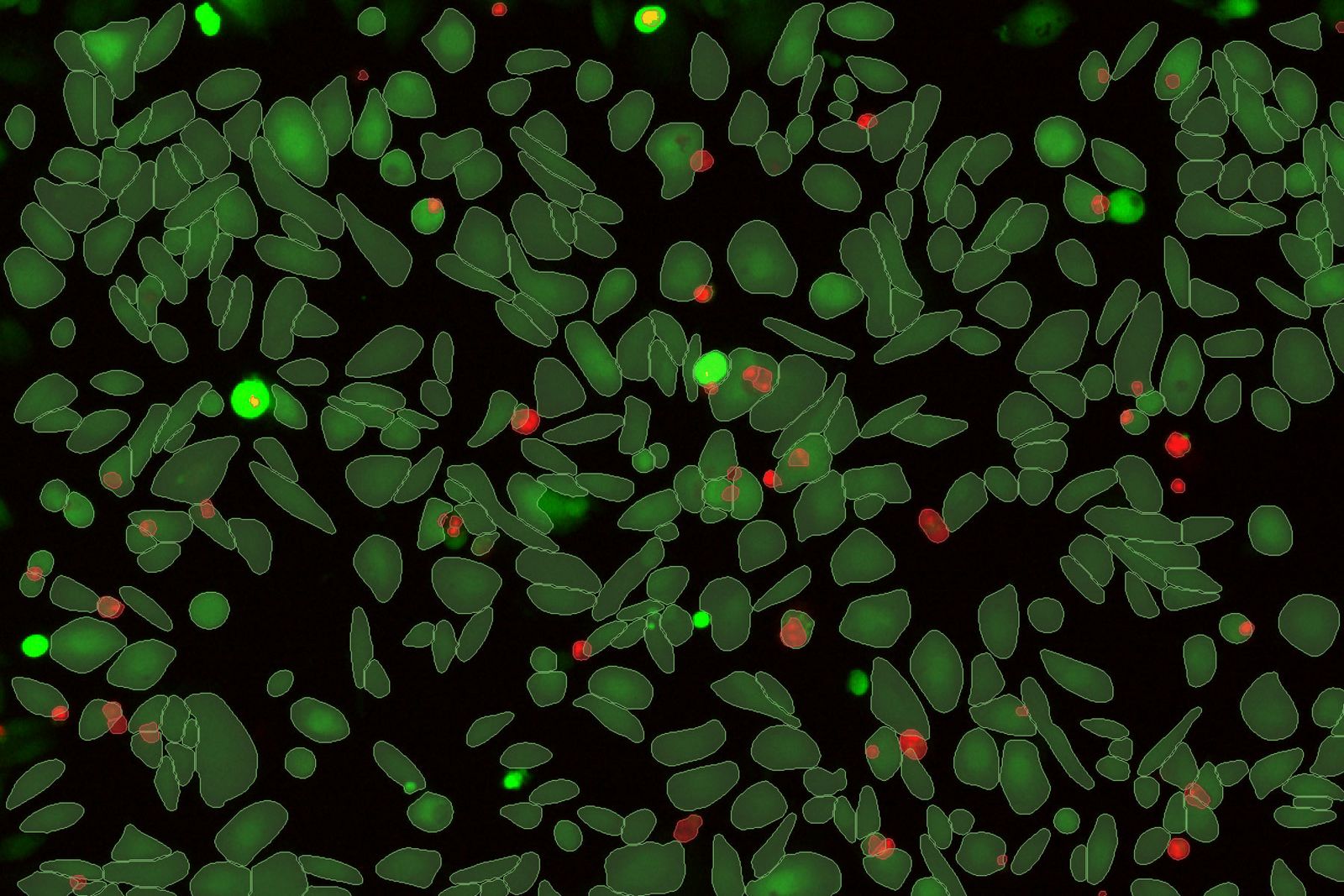
Subjectivity of analysis and poor reproducibility are key hurdles to be overcome for biological image analysis. Standard segmentation can lead to sub-standard results and require substantial manual curation which is subject to human error. Aivia changes all of that.
다중 웰 데이터를 위한 대화형 차트
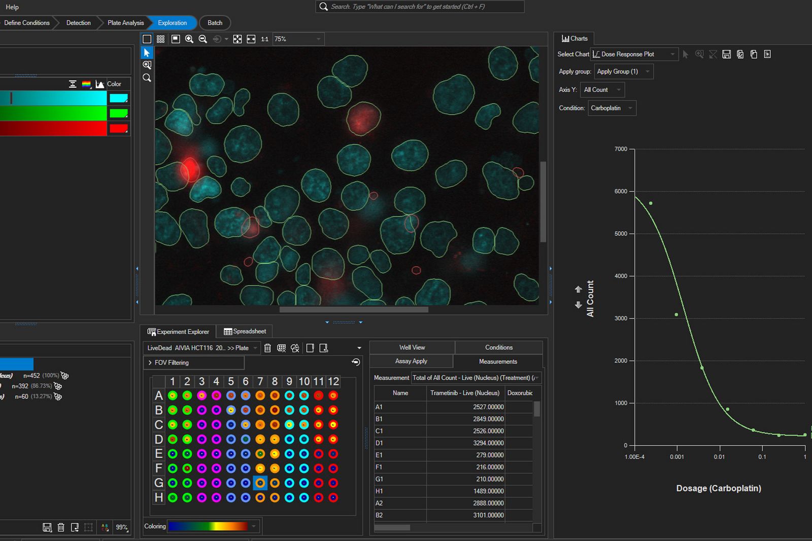
멀티웰 실험을 위한 고급 데이터 탐색. Aivia 16은 통계 분석을 위한 집계 요약 차트 및 데이터 내보내기를 통해 데이터 탐색을 강화하고 다중 웰 실험에 대한 심층적인 통찰력을 제공합니다.
- 유정별, 개체 세트별 또는 조건별로 집계된 다중 유정 데이터 세트에서 인사이트 추출하기
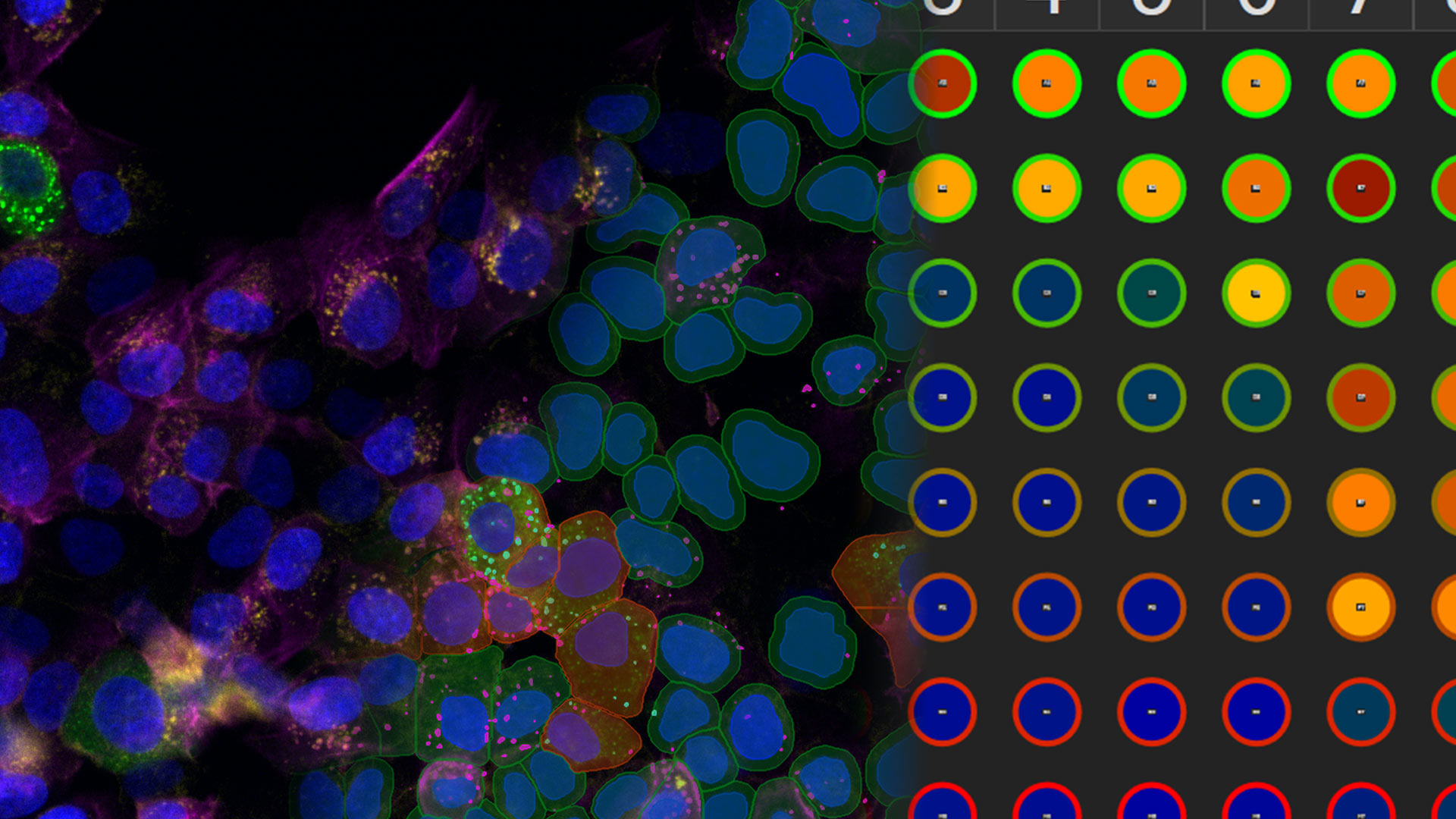
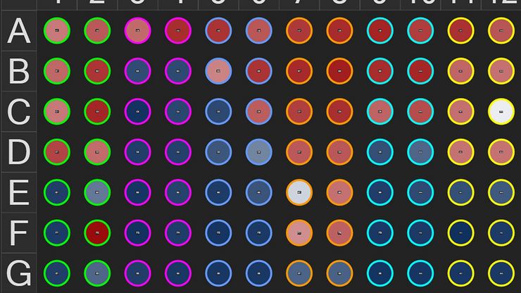
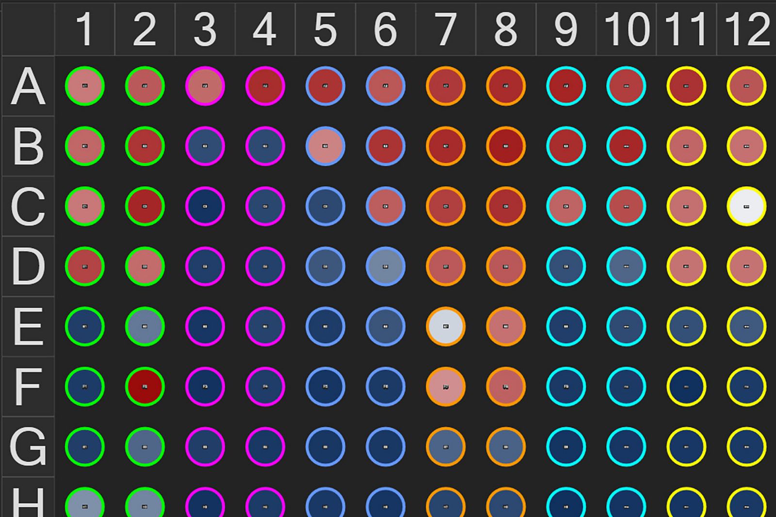
- 실험 탐색기에서 멀티웰 히트맵으로 웰별 분석 결과를 요약하고 탐색합니다
- 요약 차트에서 데이터 집계 사용자 지정
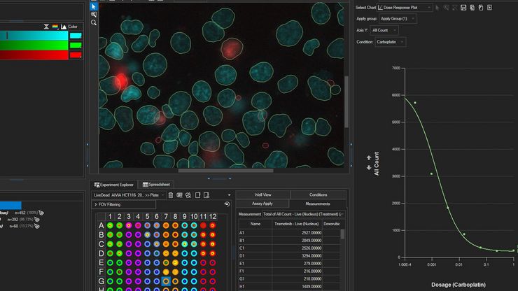
- IC50/EC50 계산을 통해 용량 반응 곡선에서 다중 웰 실험의 세포 반응을 발견하세요
- 결합된 데이터를 타사 통계 분석 소프트웨어로 내보내 결과를 더 자세히 분석할 수 있습니다
AI access for all
Aivia makes advanced data analysis accessible for all biologists - with no computer science expertise required.
The Aivia platform has been designed with the end-user in mind. This means with Aivia you can quickly and reliably generate high-quality results. The Aivia platform includes all state-of-the-art applications you will need in a unified user experience.
Quickly train laboratory users on the platform, to conduct their analysis without any specialist expertise.
- Speed up your imaging projects and publish faster
- Benefit from next-generation, easy to use machine learning segmentation and classification tools
- Conduct parameter-free image segmentation
- Easily train, update and apply deep learning models using local resources or the AiviaCloud platform
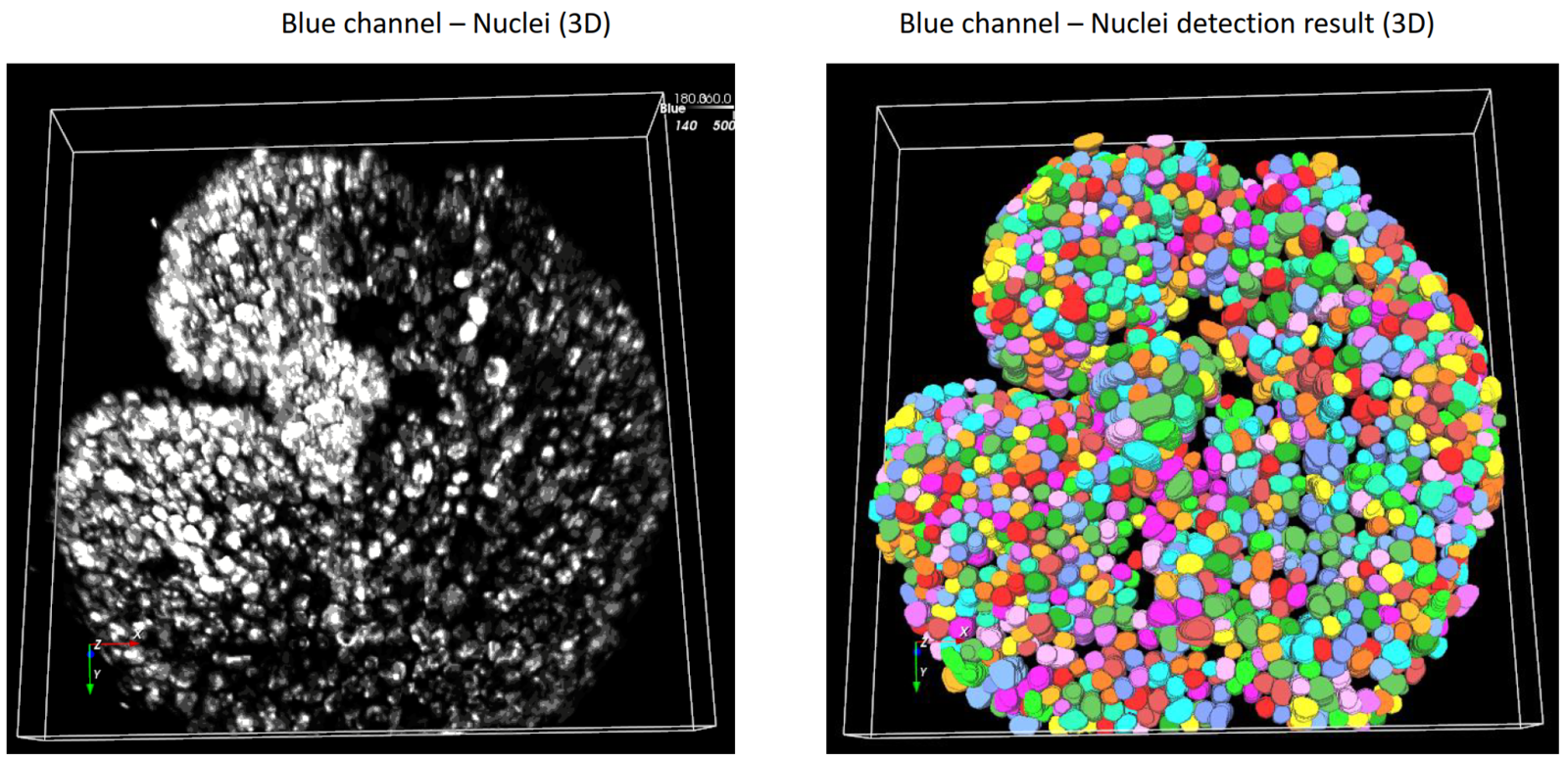
Total freedom on a single platform
Aivia's powerful and fast 2-5D visualization and analysis unlocks all the value of your data - within a single platform.
No longer does your team have to learn to operate and adopt multiple imaging and analyses systems into their workflow - the Aivia platform unifies all state-of-the-art applications you will need in a unified user experience. Aivia can leverage both local and cloud computing resources. You can install and use Aivia both on your local computer as well as via a web browser, AiviaWeb. Aivia works seamlessly with all microscopy imaging systems.
Your team can also access all files created by your imaging systems anywhere - all you need is an internet connection.
- Powerful and fast 2-5D visualization and analysis - accessible anywhere
- Includes 22 applications and 20 pre-trained deep learning models (image segmentation, restoration and virtual staining)
- Reliable and easy to use cloud access with flexible IT architectures supported
- Over 45 microscopy file formats supported

Start a free trial
Using state-of-the-art, AI-first software architecture, Aivia is a uniquely innovative and complete 2-to-5D image visualization, analysis and interpretation platform designed for the reliable processing and reconstruction of highly complex images in just minutes.
- Make AI-enhanced image analysis accessible for all - with no computer science expertise required
- Leverage machine learning capabilities to generate robust and reproducible segmentation results
- Realize powerful and fast 2-5D visualization and analysis to unleash the value of your data - all within a single platform
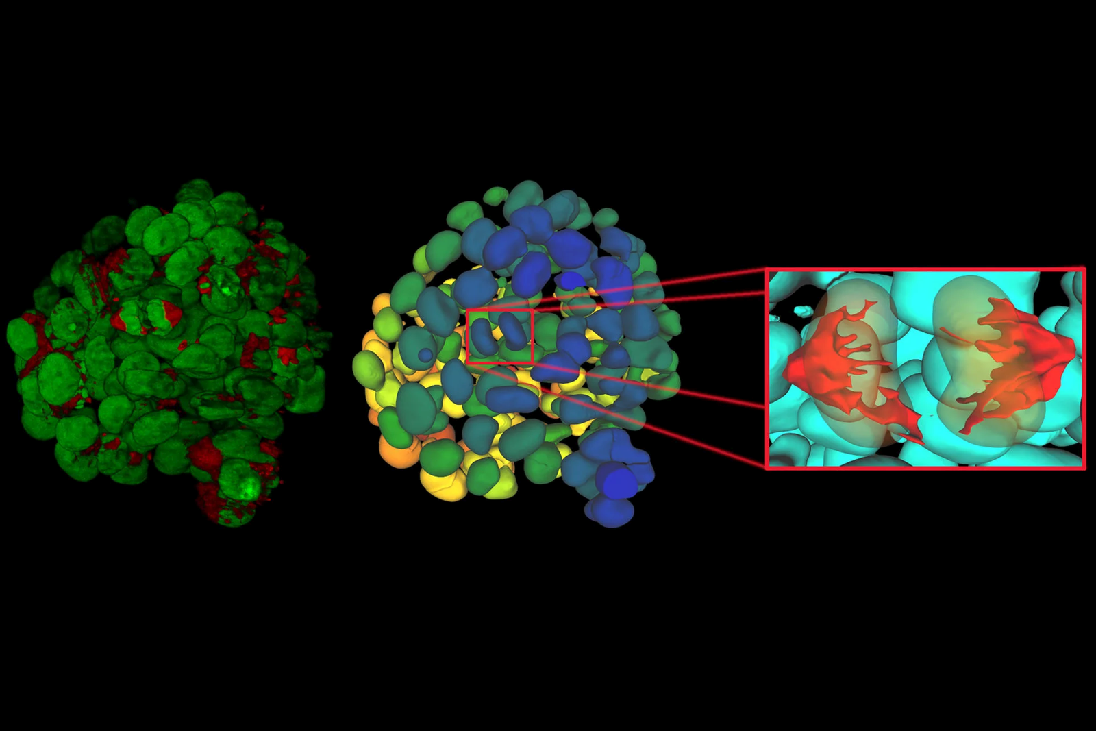
Explore Aivia subscription plans
Aivia is available via subscription, giving you the flexibility to select a plan that fits your lab’s needs now, or in the future as your research needs evolve.
With technical support and free software upgrades for the duration of your subscription, you will always have the lasted AI-powered analysis tool for your research.
- Go - The essential tools to get you started with AI image analysis
- Elevate - Take your AI image analysis to the next level with CellBio or Neuro
- Apex - A comprehensive AI image analysis platform for labs and core facilities
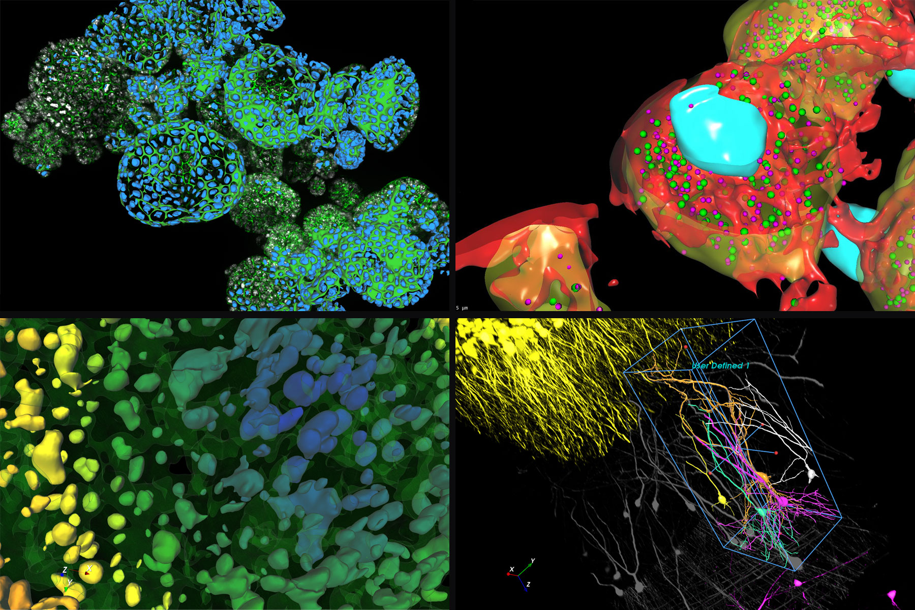
Everything you need to start analyzing images
Aivia Go offers state-of-the-art image visualization and analysis tools in a single platform, including multiple AI-powered features, to meet your challenging image visualization and analysis needs. Simple segmentation workflows and batch processing capabilities yield results faster, helping you leap from data to publication.
Aivia Go supports a large range of applications from 2D to 5D image analysis tasks such as detection and tracking. It is the ideal package for research laboratories and imaging core facilities. No computer expertise required.
- AI-powered classifiers
- 12+ image analysis recipes
- 2D -5D image visualization
Features
Image Analysis Recipes - Deploy a wide range of 2D to 5D image analysis recipes for the most popular analysis applications:
- Count cells, nuclei and particles in 2D and 3D
- Track cells (in phase contrast and fluorescence), nuclei, and particles in 2D and 3D
- Cell proliferation and wound healing assays (phase contrast)
- Neurite outgrowth
- Stem cell colony detection (phase contrast)
- Cell tracking (phase contrast)
Descubrimiento rápido de información en experimentos multipocillos complejos
Aivia Accelerate permite una rápida exploración de las relaciones entre variables clave en experimentos multipocillos complejos, permitiendo a los científicos extraer información procesable de los datos de imagen con una configuración mínima y sin necesidad de codificación.
Características
- Analice los resultados agregados en todas las condiciones experimentales utilizando gráficos de resumen con estadísticas de población
- Acelere los análisis con flujos de trabajo preconfigurados
- Despliegue de un algoritmo generalista de segmentación de células aprendizaje profundo preentrenado para la segmentación por ejemplos
- No se requiere entrenamiento de modelos, codificación ni experiencia en IA
- Clasifique automáticamente las poblaciones celulares
- Generación automática de informes exhaustivos
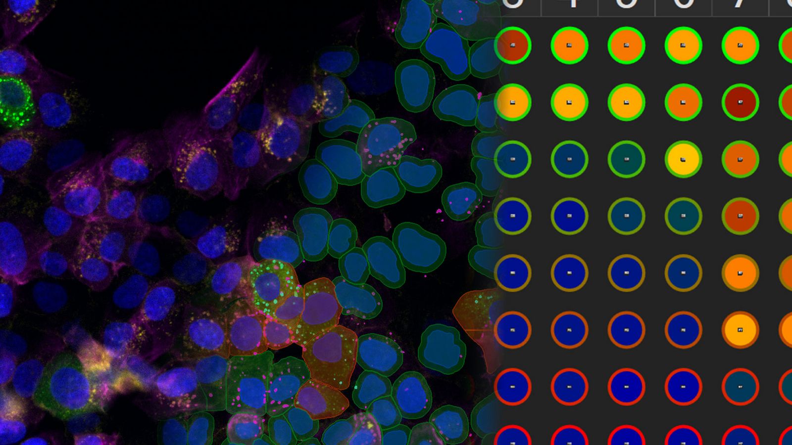
Take your AI image analysis to the next level with Aivia’s simple workflows
A complete solution for research laboratories and core imaging facilities, Aivia Elevate subscribers can choose specialized AI solutions for either neuro or cell biology image visualization and analysis.
Analyze cellular interactions and measure the distances between different compartments down to the channel level with Elevate for CellBio . It includes tools to perform accurate cell detection in 2D multiplexed images, identify cell phenotypes and explore spatial insights. Or automate challenging neuroimaging tasks, such as neuron tracing, dendrite segmentation, and more with Elevate for Neuro.
Want both options? Upgrade to Aivia Apex for full functionality plus the ability to apply your own deep learning models.
Features
Cell or Neuron Analysis Recipes - Choose between specialized analysis tools for neuroscience or cell biology research
- Neuron Analysis Recipes (Fluorescence/EM): Detect and trace neurons in 3D data sets
- Cell Analysis Recipes (2D/3D): Observe complex cellular interactions in 2D and 3D data sets
A comprehensive AI image analysis platform for labs and core facilities
Aivia Apex is a comprehensive microscopy image analysis solution for researchers who need a variety of image analysis applications. Apex also provides microscopists with the flexibility to apply their own deep learning models, third party models or open-source repositories.
Ideal for large research groups and core imaging facilities, Aivia Apex’s Floating License Manager allows a site to run all licenses on any machine on their local network.
- Everything in Elevate Neuro & CellBio
- Expanded deep learning capabilities
- Floating license manager
Features
Image Analysis Recipes - Deploy a wide range of 2D to 5D image analysis recipes for the most popular analysis applications
- Nuclei count and tracking
- Cell count and tracking
- Particle count and tracking
- Cell proliferation assay
- Neurite outgrowth
- Wound healing (phase contrast)
- Stem cell colony detection (phase contrast)
- Cell tracking (phase contrast)
- 3D Object Analysis and Tracking recipes including neuron and cell analysis
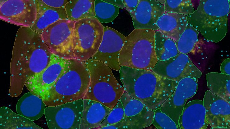
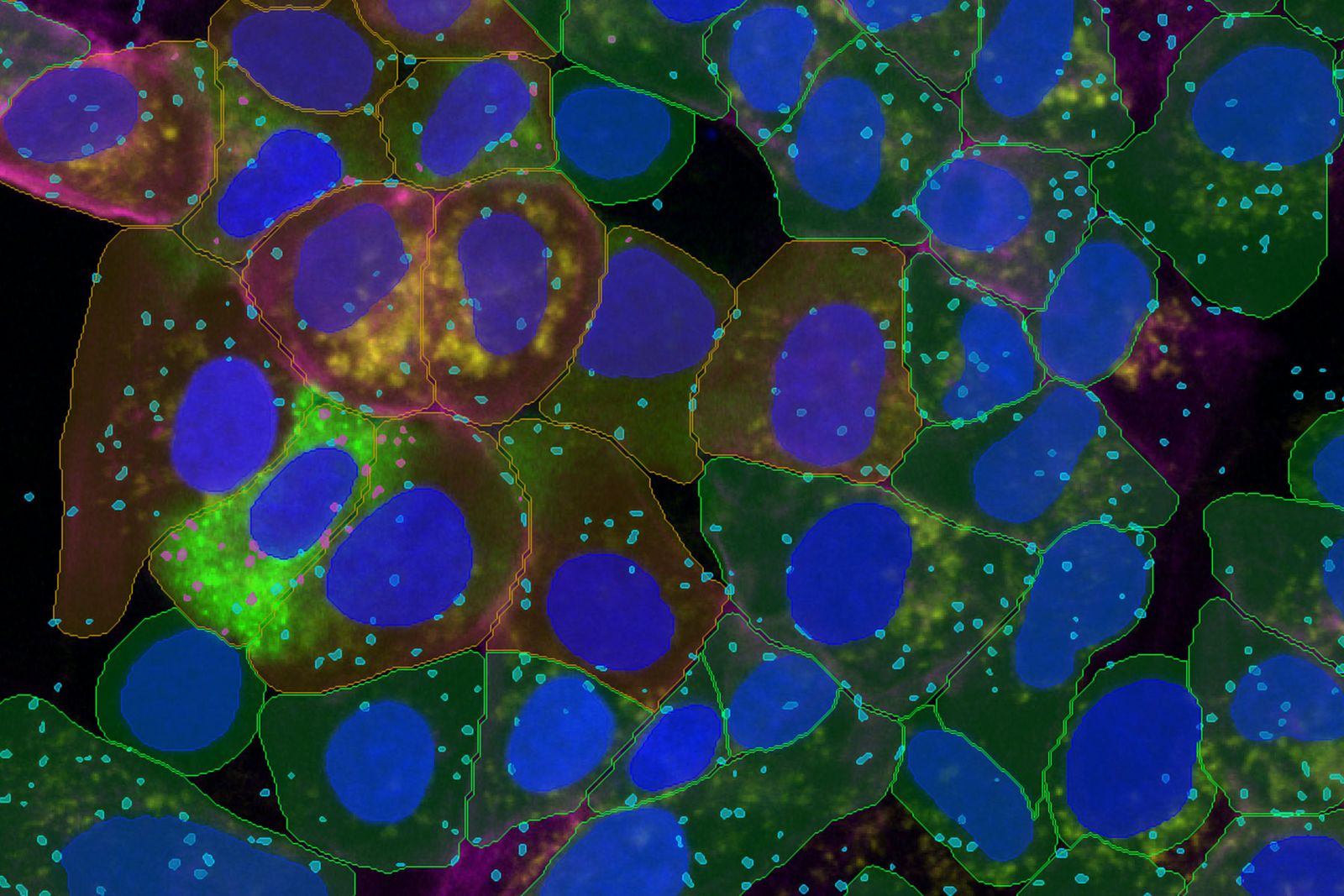
예제별 세그먼트, 한 단계 더 진화하다
더 적은 입력으로 더 빠르게 더 많은 인사이트를 얻으세요. Aivia 16의 예제별 세그먼트는 특허 출원 중인 Aivia의 딥러닝 알고리즘을 기반으로 최소한의 입력만으로도 정확한 세포 검출을 구현하여, 간편성과 정밀성의 새로운 기준을 제시합니다.
- 단 3개의 샘플만으로 2D 또는 3D 세포를 정확하게 분할합니다.
- 2D/3D 멀티플렉스 셀 감지를 위한 여러 이미지 채널 결합
- 세포, 핵 및 무제한의 소포 유형을 2D로 세분화합니다
- 한 번에 다양한 세포 집단의 표현형 분석
- 최대 12.8배 빠른 세포 및 핵 검출
다중 웰 데이터 가져오기
손쉬운 멀티웰 데이터 가져오기. Aivia 16은 연구자들이 데이터를 원활하게 가져오고, 탐색하고, 분석할 수 있도록 지원합니다. Mica, DMi8 및 STELLARIS 이미징 시스템의 LIF 또는 XELF 파일에서 직접 새로운 멀티웰 실험을 가져옵니다.
개선된 실험 탐색기에서 멀티웰 실험의 유정 및 필드 위치를 쉽게 탐색하고 Aivia에 로드된 데이터를 빠르게 검토할 수 있습니다.
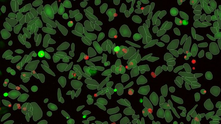
가이드 분석
간소화된 엔드투엔드 분석 워크플로. 가이드 분석은 다중 웰 샘플에서 정확성과 견고성을 높여 데이터에서 최대한의 가치를 추출하려는 사용자에게 동급 최고의 성능을 제공함으로써 발견까지의 프로세스를 간소화합니다.
- 분석 런치패드에서 분석용 가이드 시퀀스 열기
- 가이드 시퀀스는 조건 설정부터 탐지, 표현형 및 배치 처리까지 사용자를 안내합니다
- 일반적인 분석을 위한 4개의 사전 구축된 분석 가이드 시퀀스: 계수, 형태학, 생체/사멸, 세포 사멸
- 유정별, 조건별 또는 플레이트별 분석 결과 집계
- 샘플 이미지로 분석 파이프라인을 최적화하고 전체 실험에 전파하기