Upright Light Microscopes

Upright microscopes for life science research

Get the publication-quality imaging and customizable upright microscope solution you need for your Life Science research with Leica Microsystems. These powerful imaging systems feature constant color, natural light illumination, superior optics, and configurable options to provide high contrast, brilliant images for your cutting-edge biological research.

Upright microscopes for industrial and materials

Get insights into the smallest details and inspect and document results efficiently with industrial and materials upright microscopes from Leica Microsystems. Each solution can be customized with brilliant, cool LED illumination, ergonomic accessories, sophisticated digital cameras and intuitive software to meet a broad range of applications.

Need Assistance?

Contact a local imaging specialist for expert advice on the right upright microscope for your needs and budget.

Leica DM4B 6B Upright Microscopes

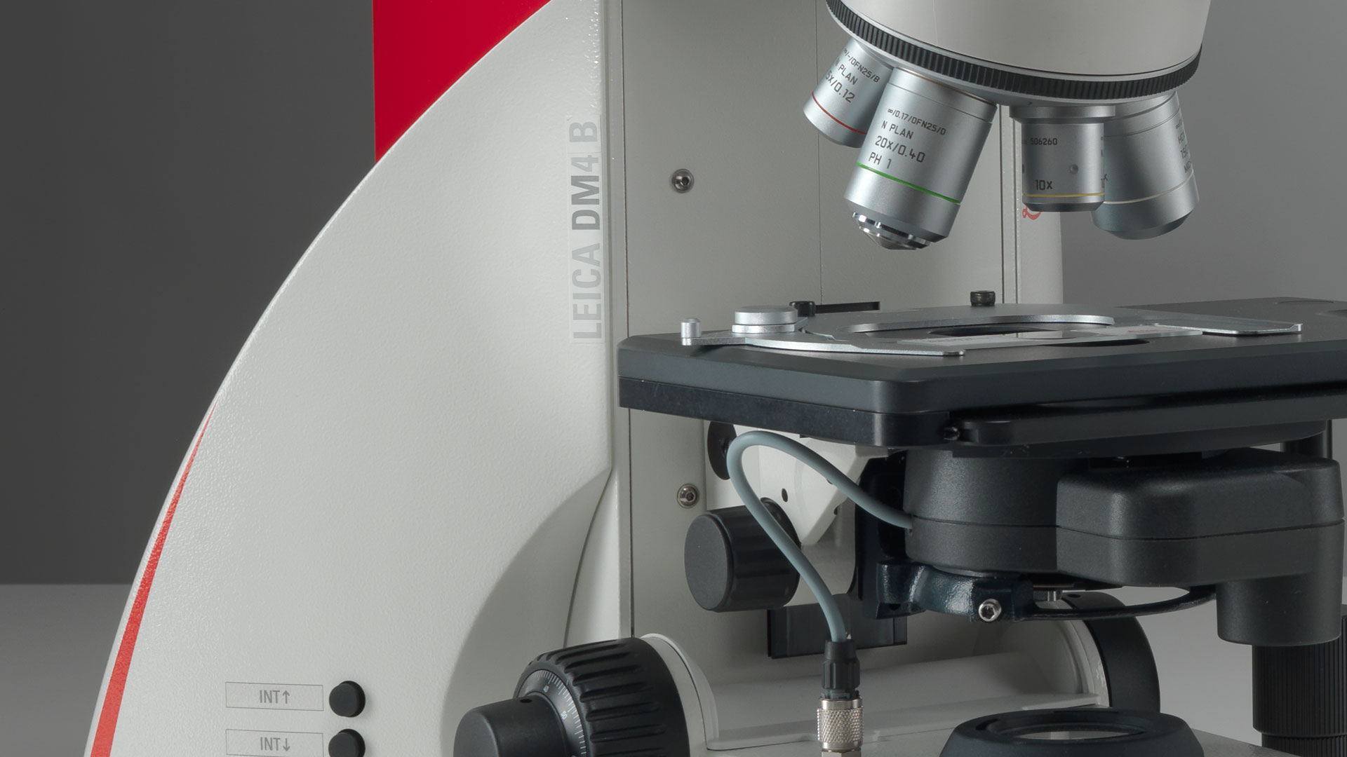
DM4 B & DM6 B Upright Microscopes

Rapid results, customized workflows, and deep insights. Optimized for imaging tissue and plant specimens, as well as pathology, these microscopes are designed to accelerate and tailor your workflows.

DM4 B & DM6 B

Follow us on Instagram
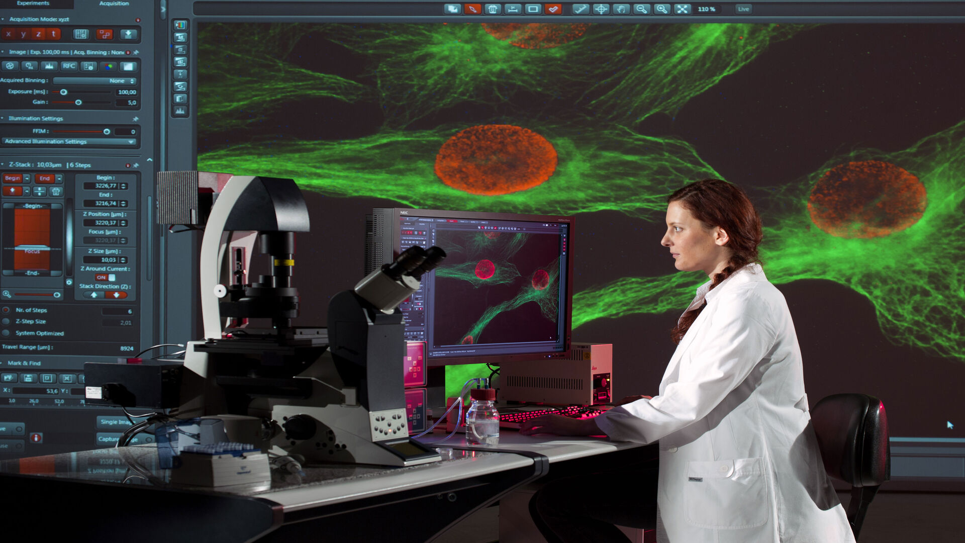
related articles

Read our latest articles about Upright Microscopes

The knowledge portal of Leica Microsystems offers scientific research and teaching material on the subjects of microscopy. The content is designed to support beginners, experienced practitioners and scientists alike in their everyday work and experiments.

More Articles

Dr. Andrew Huang, Baylor College of Medicine, in the operating room (OR) performing ear, nose and throat (ENT) surgery using the MyVeo surgical visualization headset.

A Microvascular Surgeon’s View: How MyVeo Transforms Visualization

In this article, Dr. Andrew T. Huang, MD, FACS, otolaryngologist and a head and neck reconstructive surgeon, shares how digital 3D surgical visualization with the MyVeo headset from Leica Microsystems…

A Novel Laser-Based Method for Studying Optic Nerve Regeneration

Optic nerve regeneration is a major challenge in neurobiology due to the limited self-repair capacity of the mammalian central nervous system (CNS) and the inconsistency of traditional injury models.…
Fluorescence microscopy of sectioned tissue, showing the interface between the extensor digitorum longus muscle and the common peroneal nerve in the adult rat. Regenerative peripheral nerve interface (RPNI) at 2 weeks. Image acquired using Mica. Stained for nuclei (blue), neurofilaments (green) and S100B (red). Image courtesy of Dr. Aaron Lee, Department of Bioengineering (Lab of Dr. Rylie Green), Imperial College London.

How to Image Axon Regeneration in Deep Muscle Tissue

This study highlights Dr. Aaron Lee’s research on mapping nerve regeneration in muscle grafts post-amputation. Limb loss often leads to reduced quality of life, not only from tissue loss but also due…
5 hour time-lapse maximum intensity projection of a zebrafish embryo along the z-axis at 3 days post fertilization. Left: microglia cells. Right: bright field channel. Courtesy of Prof. Francesca Peri, University of Zurich, Switzerland.

Capturing Developmental Dynamics in 3D

This application note showcases how the Viventis Deep dual-view light sheet microscope was successfully used by researchers for exploring high-resolution, long-term imaging of 3D multicellular models…
Some 2D measurements, e.g., lengths and areas, made on a PCB sample with a Leica measurement microscope using the Enersight software.

How to Select the Right Measurement Microscope

With a measurement microscope, users can measure the size and dimensions of sample features in both 2D and 3D, something crucial for inspection, QC, failure analysis, and R&D. However, choosing the…
A fruit fly (Drosophila melanogaster) observed with an Ivesta 3 stereo microscope during fly pushing (sorting of the flies). The scale bar length is 1 mm. Image courtesy of M. Benton, EMBL, Heidelberg, Germany.

A Guide to Using Microscopy for Drosophila (Fruit Fly) Research

The fruit fly, typically Drosophila melanogaster, has been used as a model organism for over a century. One reason is that many disease-related genes are shared between Drosophila and humans. It is…
These images illustrate the need for multiple z-slices to capture all gH2Ax foci in a given cell and get an accurate count.

Development and Derisking of CRISPR Therapies for Rare Diseases

This on-demand presentation by Dr. Fyodor Urnov and Dr. Sadik Kassim, originally delivered at ASGCT 2025, focused on a critical challenge in genetic medicine: how to scale CRISPR therapies from…
Example of calibrating a microscope at a higher magnification value using a stage micrometer.

Microscope Calibration for Measurements: Why and How You Should Do It

Microscope calibration ensures accurate and consistent measurements for inspection, quality control (QC), failure analysis, and research and development (R&D). Calibration steps are described in this…

Integrated Serial Sectioning and Cryo-EM Workflows for 3D Biological Imaging

This on-demand webinar explores how integrated tools can support electron microscopy workflows from sample preparation to image analysis. Experts Andreia Pinto, Adrian Boey, and Hoyin Lai present the…
SEM image of the full Li-NMC electrode sample, showing the two porous layers and the metal film at the center of the structure.

Revealing Sodium Battery Degradation via Cryo-EM and CryoFIB

Explore how cryogenic electron microscopy and focused ion beam techniques uncover the intrinsic structure of sodium battery interfaces. This webinar presents a new degradation model based on separator…

Materials Science and Inspection

Excellent sample preparation and imaging methods are key for inspecting materials and visualizing the fine details with reliability and accuracy. Inverted microscopes enable you to achieve this goal in a more efficient way compared to upright microscopes. They offer you high-quality optics and intelligent automation for optimal workflows. 

Read More

Life Science Research

Inverted microscopes are often used in life science research for the visualization, measurement, analysis, and documentation of live cell and tissue cultures. Continuous innovative technical advances for inverted microscopes support the ever-changing imaging needs of life science researchers. 

Read More

Scroll to top