
Science Lab
Science Lab
The knowledge portal of Leica Microsystems offers scientific research and teaching material on the subjects of microscopy. The content is designed to support beginners, experienced practitioners and scientists alike in their everyday work and experiments. Explore interactive tutorials and application notes, discover the basics of microscopy as well as high-end technologies – become part of the Science Lab community and share your expertise!
Filter articles
Tags
Story Type
Products
Loading...

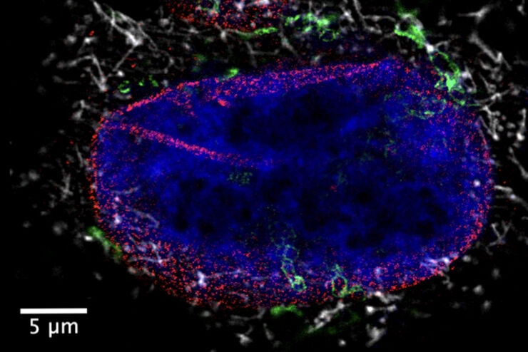
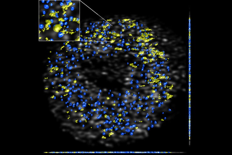
Extended Live-cell Imaging at Nanoscale Resolution
Extended live-cell imaging with TauSTED Xtend. Combined spatial and lifetime information allow super-resolution microscopy at extremely low light dose.
Loading...
, actin network (ATTO 647N), and nuclear pore basket (CF 680R).")
The Guide to STED Sample Preparation
This guide is intended to help users optimize sample preparation for stimulated emission depletion (STED) nanoscopy, specifically when using the STED microscope from Leica Microsystems. It gives an…
Loading...

Five-color FLIM-STED with One Depletion Laser
Webinar on five-color STED with a single depletion laser and fluorescence lifetime phasor separation.
Loading...
A Versatile Palette of Fluorescent Probes
Researchers at the Max Planck Institute for Medical Research in Heidelberg have developed a general strategy to synthesize live-cell compatible fluorogenic probes, and the result are the new MaP (Max…
Loading...
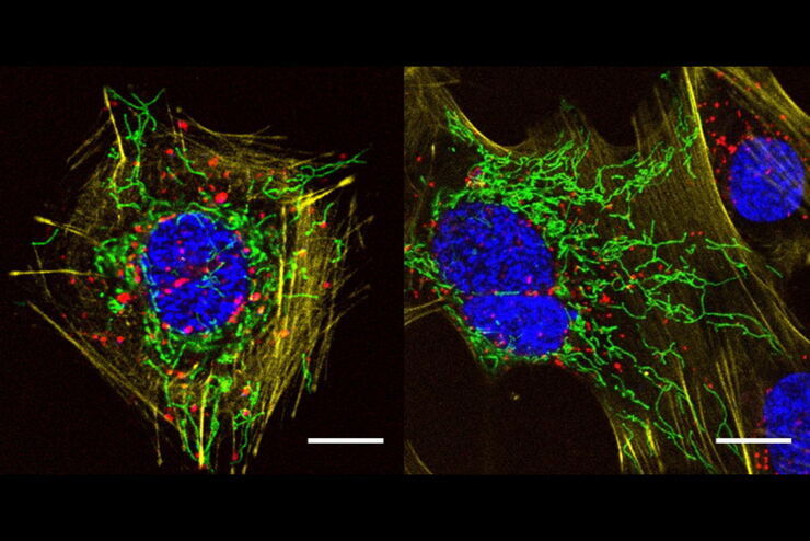
 and STED (right) imaging.")
Mitochondrial Dynamics Quantitatively Revealed by STED Nanoscopy
Mitochondria are vital subcellular organelles, which can generate energy to support the entire lifecycle of biological cells. However, conventional fluorescence microscopy cannot provide a high enough…
Loading...
Benefits of Combining STED and Lifetime
In this interview, Professor Alberto Diaspro talks about the advantages of the White Light Laser and the TauSTED capabilities of STELLARIS 8 STED. He speaks about his experience with the confocal…
Loading...
Effects of Clearing Media on Tissue Transparency and Shrinkage
This study comprehensively evaluates the effects of different clearing media on tissue transparency and shrinkage by comparing freshly dissected dipteran fly brains with their cleared equivalents.…
Loading...
DNA Replication in Cancer Cells
DNA synthesis can be impeded by collisions between the DNA replication machinery and co-transcriptional R-loops leading to a major source of genomic instability in cancer cells. In this paper we…
Loading...
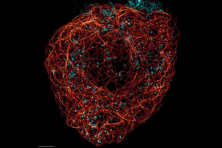
Super-Resolution Microscopy Image Gallery
Due to the diffraction limit of light, traditional confocal microscopy cannot resolve structures below ~240 nm. Super-resolution microscopy techniques, such as STED, PALM or STORM or some…