Corporate Communications

Leica Microsystems develops and manufactures microscopes and scientific instruments for the analysis of microstructures and nanostructures.
We offer scientific research and teaching material on the subjects of microscopy. The content is designed to support beginners, experienced practitioners and scientists alike in their everyday work and experiments. Explore interactive tutorials and application notes, discover the basics of microscopy as well as high-end technologies.
Follow us

Efficient Long-term Time-lapse Microscopy
When doing time-lapse microscopy experiments with spheroids, there are certain challenges which can arise. As the experiments can last for several days, prolonged sample survival must be achieved…

Dr. Tawfik Shares his Expert View on Direct Horizontal Chopping in Cataract Surgery
It is estimated that nearly 28 million cataract surgery procedures are performed worldwide every year. Phacoemulsification is the most common method used to remove the cataract and chopping maneuvers…

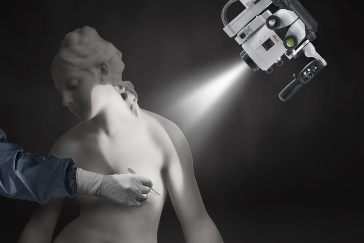
Free Flap Procedures in Oncological Reconstructive Surgery
Free flap surgery is considered the gold standard for breast, head and neck reconstructions for cancer patients. These procedures, which enable functional and aesthetic rehabilitation, can be quite…
 or Minor’s syndrome")
Minor’s Syndrome Surgical Intervention by Prof. Vincent Darrouzet
Minor’s disease, also called Superior Semicircular Canal Dehiscence (SSCD) or Minor’s syndrome, is a rare disorder of the inner ear that affects hearing and balance. The disease is characterized by…
How to Choose a Microscope for Reconstructive Surgery
Plastic and reconstructive surgery requires excellent visualization to repair intricate and fine structures. Oncological reconstructive surgery procedures are among the most delicate, including breast…

Advances in Oncological Reconstructive Surgery
Decision making and patient care in oncological reconstructive surgery have considerably evolved in recent years. New surgical assistance technologies are helping surgeons push the boundaries of what…

Accurately Analyze Fluorescent Widefield Images
The specificity of fluorescence microscopy allows researchers to accurately observe and analyze biological processes and structures quickly and easily, even when using thick or large samples. However,…
Live-Cell Imaging Techniques
The understanding of complex and/or fast cellular dynamics is an important step for exploring biological processes. Therefore, today’s life science research is increasingly focused on dynamic…
A New Method for Convenient and Efficient Multicolor Imaging
The technique combining hyperspectral unmixing and phasor analysis was developed to simplify the process of getting images from a sample labeled with multiple fluorophores. This aggregate method…
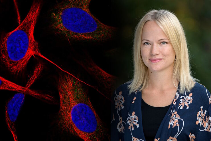
Applying AI and Machine Learning in Microscopy and Image Analysis
Prof. Emma Lundberg is a professor in cell biology proteomics at KTH Royal Institute of Technology, Sweden. She is also the director of the Cell Atlas, an integral part of the Swedish-based Human…
The AI-Powered Pixel Classifier
Achieving reproducible results manually requires expertise and is tedious work. But now there is a way to overcome these challenges by speeding up this analysis to extract the real value of the image…
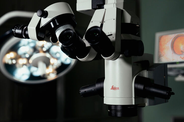
How to Select a Microscope for Cataract Surgery
What to consider in the selection of an ophthalmic microscope for cataract procedures. Bearing these aspects in mind will equip surgeons well for talks with manufacturer representatives. Many…

Intravital Microscopy of Cancer
Join our guest speaker Prof Dr Jacco van Rheenen, as he presents his work on the identity, behavior and fate of cells that drive the initiation and progression of cancer.
Save Time and Effort with AI-assisted Fluorescence Image Analysis
The powerful synergy of THUNDER and Aivia analyze fluorescence images with greater accuracy, even when using low light excitation.
Tracking Single Cells Using Deep Learning
AI-based solutions continue to gain ground in the field of microscopy. From automated object classification to virtual staining, machine and deep learning technologies are powering scientific…

Learning the Cellular Architecture from its Optical Properties
In the last 3 years, microscopists have started to use "AI based" solutions for a wide range of applications, including image acquisition optimization (smart microscopy), object classification, image…
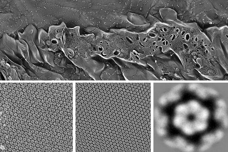
Advancing Cellular Ultrastructure Research
Freeze-fracture and freeze-etching are useful tools for studying flexible membrane-associated structures such as tight junctions or the enteric glycocalyx. Freeze-fracture and etching are two…
Benefits of Combining STED and Lifetime
In this interview, Professor Alberto Diaspro talks about the advantages of the White Light Laser and the TauSTED capabilities of STELLARIS 8 STED. He speaks about his experience with the confocal…