Ivesta 3
실체현미경 & 마크로현미경
광학 현미경
제품소개
홈
Leica Microsystems
Ivesta 3 Greenough 실체 현미경
직관적. 다용도. 신뢰성.
최신 기사를 읽어 보세요
Quality Assurance Improvement Across Industries
Precision is paramount. Imagine a pacemaker that fails mid-operation or a semiconductor flaw that causes a critical system crash. In industries, such as medical devices, electronics, and…
A Guide to C. elegans Research – Working with Nematodes
Efficient microscopy techniques for C. elegans research are outlined in this guide. As a widely used model organism with about 70% gene homology to humans, the nematode Caenorhabditis elegans (also…
A Guide to Using Microscopy for Drosophila (Fruit Fly) Research
The fruit fly, typically Drosophila melanogaster, has been used as a model organism for over a century. One reason is that many disease-related genes are shared between Drosophila and humans. It is…
Boston and San Francisco Innovation Hubs
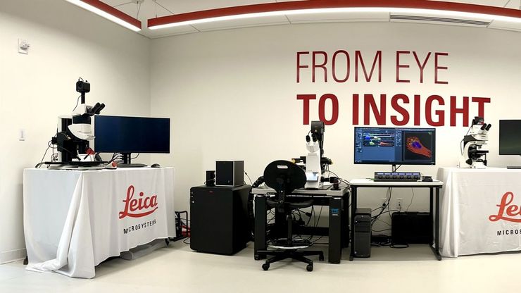
Boston and San Francisco Innovation Hubs are here to help you advance scientific discovery. We provide researchers access to state-of-the-art microscope technology and expert guidance. Located in the…
해부 현미경
해부를 해야 하는데 해부 현미경의 접안렌즈를 보는 데 더 많은 시간을 보내기도 합니다. 라이카 마이크로시스템즈에서는 다양한 종류의 현미경과 해부 현미경의 부품 및 액세서리를 선택할 수 있으므로 요건에 가장 적합한 현미경 솔루션을 찾을 수 있습니다.
Depth of Field in Microscope Images
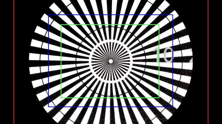
For microscopy imaging, depth of field is an important parameter when needing sharp images of sample areas with structures having significant changes in depth. In practice, depth of field is…
Understanding Clearly the Magnification of Microscopy
To help users better understand the magnification of microscopy and how to determine the useful range of magnification values for digital microscopes, this article provides helpful guidelines.
Key Factors to Consider When Selecting a Stereo Microscope
This article explains key factors that help users determine which stereo microscope solution can best meet their needs, depending on the application.
What is the FusionOptics Technology?
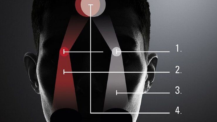
Leica stereo microscopes with FusionOptics provide optimal 3D perception. The brain merges two images, one with large depth of field and the other with high resolution, into one 3D image.
Microscope Ergonomics
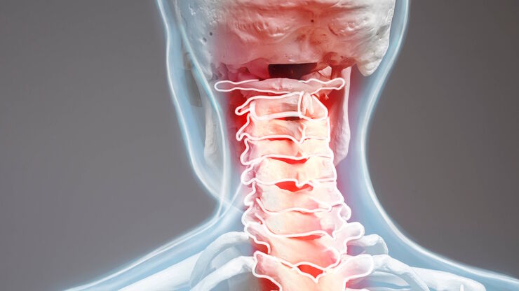
This article explains microscope ergonomics and how it helps users work in comfort, enabling consistency and efficiency. Learn how to set up the workplace to keep good posture when using a microscope.
Introduction to 21 CFR Part 11 and Related Regulations
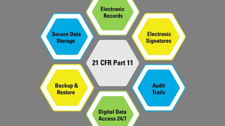
This article provides an overview of regulations and guidelines for electronic records (data entry, storage, signatures, and approvals) used in the USA (21 CFR Part 11), EU (GMP Annex 11), and China…
The History of Stereo Microscopy
This article gives an overview on the history of stereo microscopes. The development and evolution from handcrafted instruments (late 16th to mid-18th century) to mass produced ones the last 150…
Assembly & Rework Microscopes
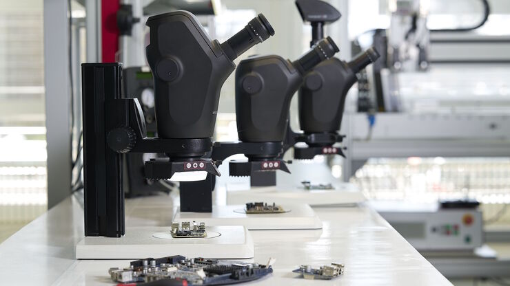
Discover Leica assembly microscopes with FusionOptics for precision rework and assembly. Tailored for electronics, automotive, medical devices, and watchmaking production needs.
Fields of Application
검사 현미경
라이카마이크로시스템즈는 다양한 산업 분야를 위한 검사 현미경과 각종 액세서리를 제공합니다. 당사의 전문가가 최적의 솔루션을 찾을 수 있도록 도와드립니다.
전자 및 반도체 산업
전자 및 반도체의 경우 효율적인 검사, 단면 및 청정도 분석, PCB, 웨이퍼, IC 칩 및 배터리의 R&D를 지원하는 솔루션이 중요합니다.
자동차 분야
Leica는 최적의 이미징 솔루션을 제공하는 신뢰할 수 있는 파트너가 되어 고객이 경쟁에서 앞서 나갈 수 있기를 희망합니다.
의료 기기 QA & QC 현미경 솔루션
효율적이고 편안한 의료 기기 검사 및 품질 관리를 위한 라이카 현미경 솔루션을 확인해 보세요.
측정 현미경
측정 현미경은 품질 관리(QC), 고장 분석 및 연구 개발(R&D) 과정에서 시료와 피처의 치수를 결정하는 데 유용합니다. 라이카 측정 현미경에 대해 자세히 알아보세요.
시계 제조업
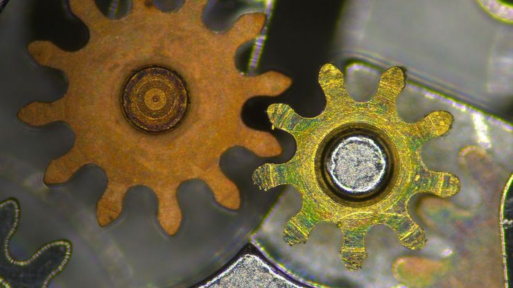
시계 제조업자와 시계 제조 산업에 있어서, Leica 실체 현미경의 높은 정밀도는 정교하게 이뤄지는 시계 조립과 높은 품질과 기술력을 보증하기 위한 신뢰할 수 있는 검사 과정을 용이하게 합니다. 저희의 인체공학적 액세서리를 사용하면, 현미경으로 장시간을 작업해도 피로를 줄일 수 있도록 고객의 필요에 따라 기기를 최적으로 구성할 수 있습니다.
교육
현미경수업은 학생들에게 가장 인기있는 과목 중 하나입니다. 우리의 교육용 현미경은 학생들의 잠재력을 최대한 발휘 할 수 있도록 도와 줄 것입니다. 실용적이고 다루기 쉽고 학생들의 관심을 끌 수 있는 이미지를 제공합니다.


