Microscope Cameras

Microscope Cameras

Microscope Cameras

Leica microscope cameras are remarkable for their fast live image speeds, short reaction times, high pixel resolution, and clear contrast. They can be installed on many of the Leica microscopes and macroscopes.

Read our latest articles about Microscope Cameras

The knowledge portal of Leica Microsystems offers scientific research and teaching material on the subjects of microscopy. The content is designed to support beginners, experienced practitioners and scientists alike in their everyday work and experiments.

More Articles

Clinical Microscopy: Considerations on Camera Selection

The need for images in pathology laboratories has significantly increased over the past few years, be it in histopathology, cytology, hematology, clinical microbiology, or other applications. They serve many purposes on top of the documentation of the diagnosis. Yet, the view through the eyepieces and the image are different in nature, the one is an optical image, the other a digital image. Looking at a few aspects of this process that are related cameras will help you make sure you can obtain the images with all the detail and color fidelity you need.

Technical Terms for Digital Microscope Cameras and Image Analysis

Learn more about the basic principles behind digital microscope camera technologies, how digital cameras work, and take advantage of a reference list of technical terms from this article.

Understanding Clearly the Magnification of Microscopy

To help users better understand the magnification of microscopy and how to determine the useful range of magnification values for digital microscopes, this article provides helpful guidelines.

Life Science Research: Which Microscope Camera is Right for You?

Deciding which microscope camera best fits your experimental needs can be daunting. This guide presents the key factors to consider when selecting the right camera for your life science research.

Image Gallery: THUNDER Imager

To help you answer important scientific questions, THUNDER Imagers eliminate the out-of-focus blur that clouds the view of thick samples when using camera-based fluorescence microscopes. They achieve this using Computational Clearing our new opto-digital technology. The result is high-speed, high-quality imaging of a large diversity of three-dimensional samples, including model organisms, tissue sections, and 3D cell cultures. Take a look at these images to see how THUNDER Imagers are already helping researchers to reveal the finest structural details even deep within a sample.

Factors to Consider When Selecting a Research Microscope

An optical microscope is often one of the central devices in a life-science research lab. It can be used for various applications which shed light on many scientific questions. Thereby the configuration and features of the microscope are crucial for its application coverage, ranging from brightfield through fluorescence microscopy to live-cell imaging. This article provides a brief overview of the relevant microscope features and wraps up the key questions one should consider when selecting a research microscope.

THUNDER Imagers: High Performance, Versatility and Ease-of-Use for your Everyday Imaging Workflows

This webinar will showcase the versatility and performance of THUNDER Imagers in many different life science applications: from counting nuclei in retina sections and RNA molecules in cancer tissue sections to monitoring calcium waves in Arabidopsis seedlings and much more.

Digital Classroom Options

As teachers, you know your big challenge is to catch and keep the students’ attention and the best chance for this is by making the environment interactive. In the case of the Microscopy Classroom, we can create an interactive environment using digital imaging components such as cameras, apps, and software. This webinar will familiarize you with the various methods you and your students can use to connect to a microscope camera and increase the learning time while enhancing the learning experience.

Imaging Live Cells

When studying live cells, common obstacles include phototoxicity and photodamage. Live‐cell microscopy often requires a compromise between image quality and cell health. Leica imaging solutions overcome these challenges for live-cell imaging, enabling the discovery of new information concerning cellular physiology and dynamics.

Key Questions

1What are Your Application Needs?

The main factor to consider when choosing a microscope camera which will best meet your needs are the applications for which it will be used. Additionally, the optical performance of your microscope is important for determining which camera can ultimately provide the best imaging results.

2What is more Important: Pixels or Resolution?

For microscopy applications, the camera with the most pixels is not necessarily the best one to use. A key criterion for microscope resolution is the numerical aperture (NA), i.e., the light-gathering power, of the optics. However, for images to have a good resolution, which reveal the fine details, at low magnifications, then you need a digital camera with a small pixel size, i.e., a sensor with a large number of pixels.

3Do You Need Enhanced Camera Performance?

Other camera features to consider for enhanced performance are: i) fast live-imaging speeds (high frames per second) for fast on-screen focusing and sample positioning; ii) large dynamic range for maximum detail in light and dark areas of the sample; and iii) true-color reproduction where you see the sample with the same colors in the camera image and via the microscope eyepieces.

Microscope Cameras Products 9

Color CMOS Microscope Camera for fast color and fluorescence tissue imaging K7

K7

Color CMOS Microscope Camera for fast color and fluorescence tissue imaging
Leica Flexacam c5 & i5 cutout

Flexacam c5 & i5

With Flexacam microscope cameras, you can see the fine sample details you need to see with precise colors and high resolution. Viewing, documenting, and sharing microscope images of your samples the...
Microscope Camera K8

Microscope Cameras

Microscope cameras from Leica are particularly remarkable for their fast live images, short reaction times, high resolution and clear contrast. And they are compatible with almost all Leica microscopes and macroscopes.

Sophisticated Microscope Cameras

We have the right camera for every application to record exactly what you see through our high-performance instruments. Photographs with a resolution of more than 12 million pixels, ultra high sensitivity and optimum color fidelity are possible with our Microscope Cameras

Imaging and Analysis of the Finest Details

Our product range covers digital Microscope Cameras with intuitive software for archiving, measurement, analysis and presentation. 100% reproducibility of the exposures and highly convenient remote control of the cameras and ensure a fast and economical workflow.

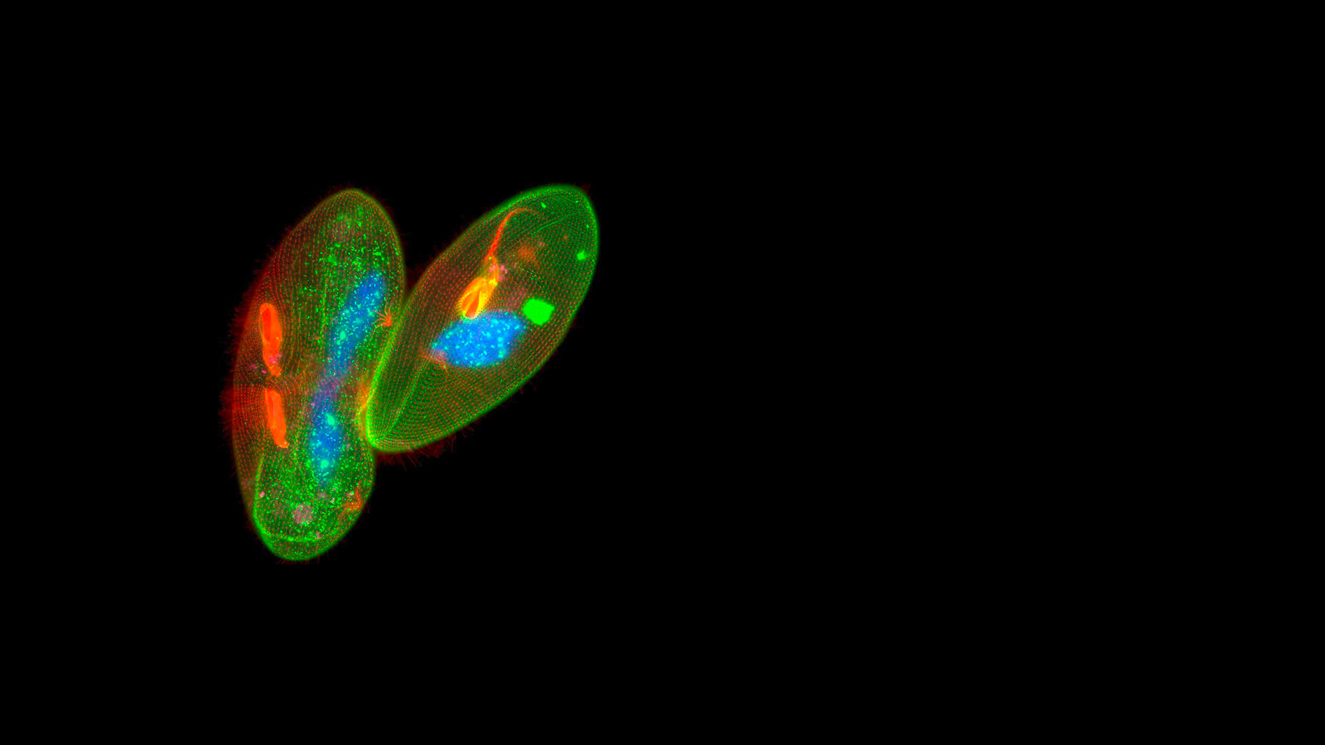
Microscope Cameras for Fluorescence Microscopy

Insights into the structures and dynamics of life

Fluorescence microscopy is a vital technique for studying biological processes. Fluorescence can be used to visualize specific subcellular structures and reveal connections between dynamic processes in live cells and tissues. A broad range of advanced live-cell imaging techniques can be employed by researchers to probe the complexities of cellular processes. Fluorescence imaging is central to many of these imaging techniques. 


To obtain excellent fluorescence images, you need a highly sensitive camera delivering a high signal-to-noise ratio and a large dynamic range resulting in a crisp fluorescence signal. Also, live-cell imaging often requires a high acquisition speed to capture fast dynamic processes. 

Why Use Fluorescence Cameras from Leica Microsystems?

The Leica fluorescence cameras are based on highly sensitive sCMOS or CCD sensors that are ideally suited for low light applications and can detect even weak signals. One camera is passively cooled to help reduce noise. The sensors also have high quantum efficiency so that you can reduce exposure times, protecting your samples from photodamage.

Monochrome and Color Microscope Cameras

Our monochrome and color cameras are designed for a broad range of applications, from basic documentation tasks to advanced live-cell imaging applications. Our Leica Application Suite (LAS) X software seamlessly integrates our cameras into the microscope system, supporting high-speed triggering.

Microscope Camera C3 close up

Microscope Cameras for Life Science

Facilitating insights into life

Life science research has a broad range of imaging needs that require solutions which address a wide variety of different applications. Microscopes cameras are vital for many life science imaging workflows like monitoring cell cultures, documenting stained specimens during morphological examinations, live-cell imaging, and the analytical methods FRAP and FRET. They generate reliable, reproducible, and quantifiable data that are essential for analysis and comparison.

For fluorescence applications, cameras should capture as many photons emitted from the sample as possible while introducing minimal additional noise to the data. Leica Microsystems offers a range of color or monochrome fluorescence cameras using sCMOS or CCD sensors.

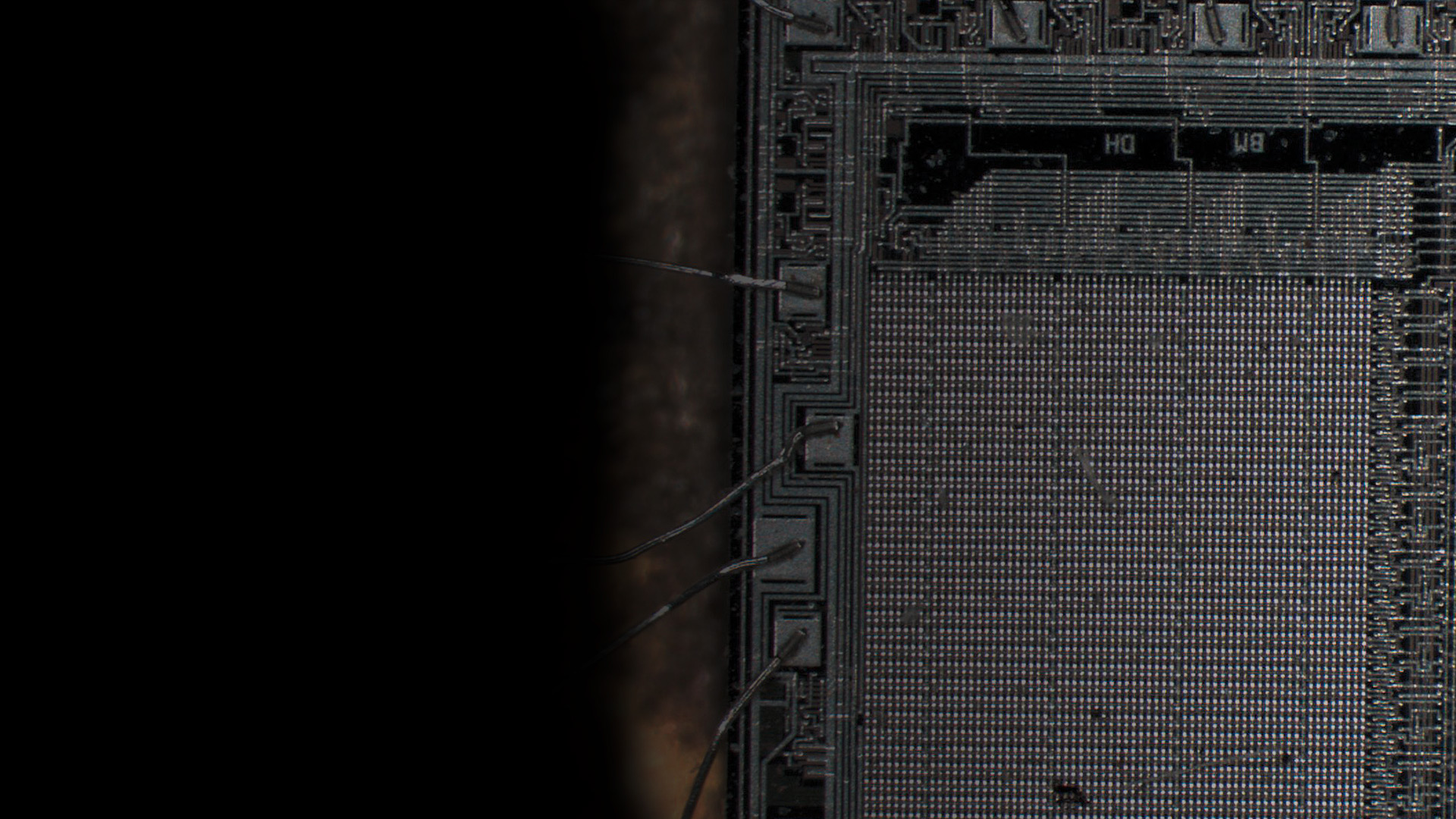
Microscope Cameras for Industry

Optimum visualization and ease-of-use

Industrial applications require microscope cameras which: i) are capable of high frame rates for fast live images to accommodate a rapid workflow; ii) can provide excellent image quality for precise analysis; and iii) are easy to use even for inexperienced users.

As a leader in microscopy innovation, Leica Microsystems provides high-performance digital microscope cameras for PC-based or stand-alone systems. The Leica portfolio includes various color cameras with CMOS- or CCD-based sensors having up to 20 MPs.

Rating Steel Quality

Foreign substances in steel, known as non-metallic inclusions, disrupt the homogeneity of the microstructure. They can have considerable influence on the steel’s mechanical properties, affecting performance and lifetime. Therefore, rating the inclusions is important for evaluating steel quality.

Read More

Education

Microscopy courses are most enjoyable when both teachers and students can focus on their subject. Our educational microscopes help you to unlock your students’ full potential: They are practical, easy to handle, and provide images that will hold your students’ attention.

Read More

Frequently Asked Questions Microscope Cameras

A camera which is installed directly onto a microscope to observe live images of samples and record them at various magnification values.

It can be operated either via connection with a computer having the appropriate software installed on it or in stand-alone mode with the image displayed directly on a monitor.

Leica microscope cameras can have from 1 to 20 megapixels. In terms of imaging resolution, it is the pixel size that is important. However, smaller pixels generally mean a greater number of megapixels on a sensor, depending on the sensor size. For more information, refer to the following articles: Introduction to Digital Camera Technology, Digital Cameras, Definitions of Basic Technical Terms for Digital Microscope Cameras and Image Analysis, What Does 30,000:1 Magnification Really Mean?

The camera is normally installed onto the microscope with a C-mount lens, but in some cases it is placed directly between the optics carrier and eyepieces.

Latest News

New Ivesta 3 stereo microscopes and Flexacam cameras extend Enersight- software-based portfolio.

Microscope Camera K3C & K3M

Suitable for a wide range of brightfield and entry-level fluorescence quantitative imaging applications in life science and industry

The new Flexacam C3 microscope camera

Interested to know more?
Talk to our experts.

Do you prefer personal consulting? Show local contacts

Scroll to top