Introduction
The inability of axons to regenerate and reinnervate after trauma and disease, such as spinal cord injury (SCI), stroke, traumatic brain injury (TBI), or multiple sclerosis (MS), results in a devastating prognosis for patients [1-3]. Numerous studies have identified two broad classes of axon growth inhibitor (AGI) proteins which are responsible for axon growth arrest [1]. These are, namely, myelin associated inhibitors (Nogo, MAG, OMgp) and the Chondroitin Sulfate Proteoglycans (CSPGs). Experiments that negate the activity of these inhibitors in vivo have shown a slight increase in regeneration of damaged axons, but a more dramatic restitution of function [2]. An alternative hypothesis to “long-distance” axon regeneration-mediated restitution of function would be the reorganization of intact spinal circuitry that often remains after SCI [3]. One of the goals of such experiments is to evaluate the potential for intact spinal circuits to replace lost connections and further to define whether negating the actions of AGIs supports adaptive or maladaptive axonal reorganization.
In this study, both widefield microscopy and the THUNDER imaging technology were used. The goal was to see if there is a difference in efficiency for screening of active versus non-active axons.
Methods
Specimen
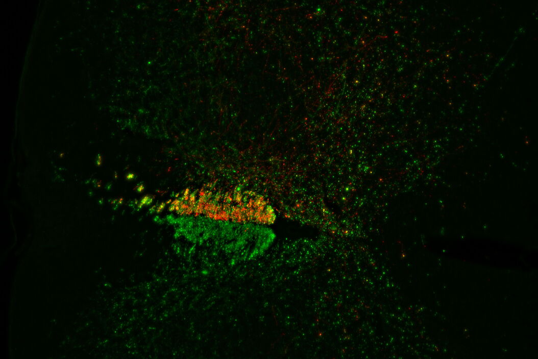
A mouse model organism was used for the study. Fluorescently labeled mouse spinal cord sections were harvested. Counting active axons in areas treated with injections that negate the actions of AGI’s was used to determine the efficacy of the experimental treatment.
Imaging
Image data were acquired using a THUNDER Imager 3D Tissue from Leica Microsystems. Stacks of 10 Z-planes were taken with a PL APO 10X/0.45 objective and a DFC9000 GT sCMOS camera.
Results
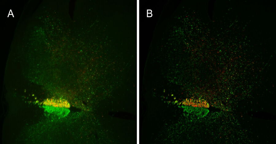
Images of a mouse spinal cord section are shown in figure 1. The left image (Fig. 1A) is the raw widefield fluorescence image shown as a maximum intensity projection (MIP). The right image (Fig. 1B) is a MIP of the same data processed with Large Volume Instant Computational Clearing (LVICC) used in the THUNDER technology. The disconnected, inactive axons fluoresce green while the re-connected, active ones fluoresce red.
Conclusion - Faster Axon Screening with THUNDER Imaging
The results (Fig. 1) show that a better discrimination of the location and number of red (active) versus green (inactive) fluorescing axons is possible with THUNDER images compared to standard widefield images. Based on this result, THUNDER imaging could allow a faster screening of the axons, enabling the effectiveness of experimental treatments to be determined more efficiently. Those treatments found to be the most worthwhile can be further investigated as potential ways to counter the effects of AGI proteins after brain or spinal cord trauma or disease.
Acknowledgements
Image data is courtesy of Prof. William Cafferty, Neurology and Neuroscience, School of Medicine, Yale University, New Haven, CT, USA.
References
- Y. Zou, M. Stagi, X. Wang, K. Yigitkanli, C.S. Siegel, F. Nakatsu, W.B.J. Cafferty, S.M. Strittmatter, Gene-Silencing Screen for Mammalian Axon Regeneration Identifies Inpp5f (Sac2) as an Endogenous Suppressor of Repair after Spinal Cord Injury, Journal of Neuroscience (2015) vol. 35, is. 29, pp. 10429-10439, DOI: 10.1523/JNEUROSCI.1718-15.2015
- K.L. Fink, S.M. Strittmatter, W.B.J. Cafferty, Comprehensive Corticospinal Labeling with mu-crystallin Transgene Reveals Axon Regeneration after Spinal Cord Trauma in ngr1−/− Mice, Journal of Neuroscience (2015) vol. 35, iss. 46, pp. 15403-15418, DOI: 10.1523/JNEUROSCI.3165-15.2015
- K.L. Fink, W.B.J. Cafferty, Reorganization of Intact Descending Motor Circuits to Replace Lost Connections After Injury, Neurotherapeutics (2016) vol. 13, iss. 2, pp. 370–381, DOI: 10.1007/s13311-016-0422-x
Related Articles
-
 imaged with the THUNDER Imager 3D Cell Culture. Courtesy of Dr. F.T. Arroso Martins, Tamere University, Finland.")
How to Get Deeper Insights into your Organoid and Spheroid Models
In this eBook, learn about key considerations for imaging 3D cultures, such as organoids and…
Nov 22, 2023Read article -
 acquired using THUNDER Imager Live Cell. Image courtesy of Janina Kaspar and Irene Santisteban, Schäfer Lab, TUM.")
Imaging Organoid Models to Investigate Brain Health
Imaging human brain organoid models to study the phenotypes of specialized brain cells called…
Jul 11, 2023Read article -
. Image courtesy of Prof. Hui Guo, School of Life Sciences, Central South University, China")
How Microscopy Helps the Study of Mechanoceptive and Synaptic Pathways
In this podcast, Dr Langenhan explains how microscopy helps his team to study mechanoceptive and…
Jun 22, 2023Read article