 tissue.")
Unlocking colon cancer's hidden spatial mysteries
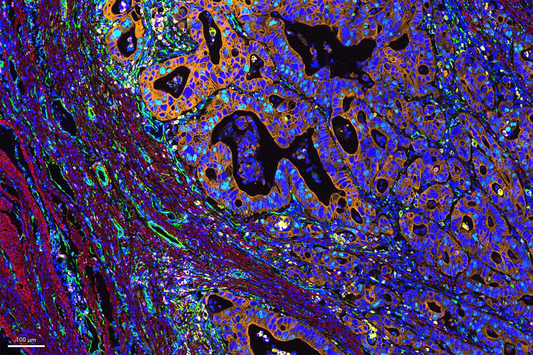
Colorectal cancer is a high burden cancer in the United States, leading to the second highest cause of cancer deaths. Despite chemotherapeutic intervention and surgical resection, recurrent disease may occur in some patients and typically, has a poorer prognosis. Thus, there is a demand for advanced therapies such as immunotherapies for improved clinical outcomes. An in-depth understanding of the colon cancer microenvironment is necessary to improve outcome in this subset of patients. Spatial biology approaches are well positioned to comprehensively uncover the molecular and biological mechanisms of cancer cell aggression in colorectal cancer. Here, we visualized nearly 30 biomarkers in colon adenocarcinoma (CAC) tissue using the Cell DIVE multiplexed imaging workflow and validated antibodies from Cell Signaling Technology (CST®). Using this approach, we were able to probe multiple pathways of interest in cancer progression, such as vascularization of tumor tissue, immune cell responses, and cell proliferation. Together, these data allow the creation of a spatial map of tumor cell aggression and offer predictive value for the progression of the disease. AI-driven analysis with Aivia allows the precise and automated cell segmentation and phenotyping necessary for the individual discrimination of cell identity. With automated clustering analysis within Aivia, hidden spatial discoveries are accessible in an unbiased way. Deep molecular and architectural insights on this level are possible only with a spatial biology approach and offer multiple downstream hypotheses to further understand cancer progression and interrogate the heterogeneity within the tumor microenvironment.
Enabling comprehensive multiplexed imaging
Cell DIVE Multiplex Imaging Solution allows probing and imaging of dozens of biomarkers on a whole single tissue section with an iterative staining and dye inactivation workflow. At its core, Cell DIVE is a precise and adaptable open multiplexing solution that enables flexibility in antibody selection of biomarker panels used in a multiplexed imaging study. Cell Signaling Technology (CST) has a broad portfolio of IHC-validated antibodies to detect key proteins in the TME, enabling immune cell detection and phenotyping in tissue. CST offers off the shelf (OTS), ready-to-ship antibody conjugates that have been verified to work on Cell DIVE and offers custom conjugation of IHC-validated antibodies to fluorophores and other detection reagents. CST employs a rigorous approach to IHC validation, followed by verification on the Cell DIVE platform to ensure successful detection of proteins.
Related Articles
-

A Meta-cancer Analysis of the Tumor Spatial Microenvironment
Learn how clustering analysis of Cell DIVE datasets in Aivia can be used to understand…
Apr 26, 2024Read article -

Spatial Architecture of Tumor and Immune Cells in Tumor Tissues
Dig deep into the spatial biology of cancer progression and mouse immune-oncology in this poster,…
Apr 26, 2024Read article -

IBEX, Cell DIVE, and RNA-Seq: A Multi-omics Approach to Follicular Lymphoma
In a recent study by Radtke et al., a multi-omics spatial biology approach helps shed light on early…
Apr 08, 2024Read article