
Science Lab
Science Lab
The knowledge portal of Leica Microsystems offers scientific research and teaching material on the subjects of microscopy. The content is designed to support beginners, experienced practitioners and scientists alike in their everyday work and experiments. Explore interactive tutorials and application notes, discover the basics of microscopy as well as high-end technologies – become part of the Science Lab community and share your expertise!
Filter articles
Tags
Story Type
Products
Loading...

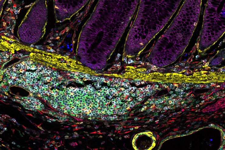
Methods to Improve Reproducibility in Spatial Biology Research
Establish reproducibility results for a Cell DIVE multiplexed imaging study in cancer research using the BAB 200 automated system from ASLS and validated antibodies from CST
Loading...
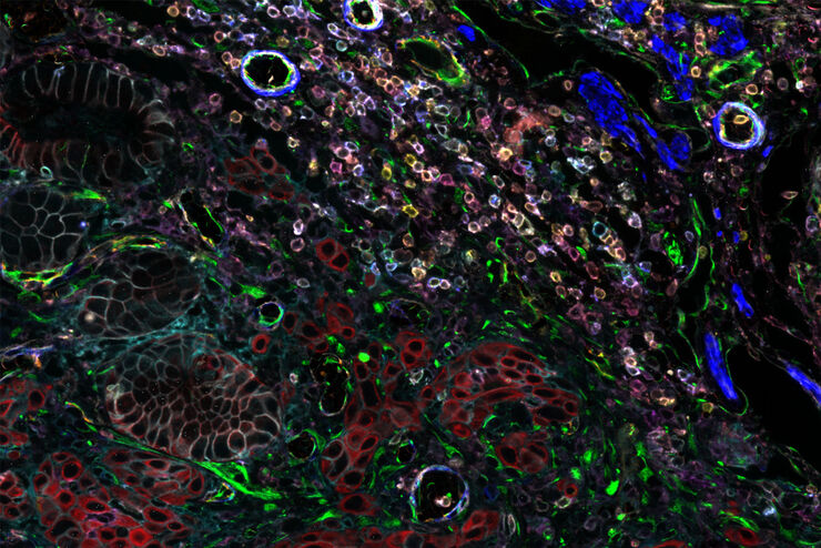
 tissue on a single slide.")
Characterizing tumor environment to reveal insights and spatial resolution
Antibodies from Cell Signaling Technology are validated for use with the Cell DIVE multiplexing workflow and used to probe cell lineages in the tumor microenvironment
Loading...

The Role of Iron Metabolism in Cancer Progression
Iron metabolism plays a role in cancer development and progression, and modulates the immune response. Understanding how iron influences cancer and the immune system can aid the development of new…
Loading...

Multiplexed Imaging Types, Benefits and Applications
Multiplexed imaging is an emerging and exciting way to extract information from human tissue samples by visualizing many more biomarkers than traditional microscopy. By observing many biomarkers…
Loading...
 and THUNDER (right) image of Ewing Sarcoma cells (SK-ES-1)")
Visualizing the Mitotic Spindle in Cancer Cells
This article demonstrates how this research is aided by visualizing more details of mitotic spindles in Ewing Sarcoma cells using the THUNDER Imager Tissue and Large Volume Computational Clearing…
Loading...
 enhanced with Aivia’s Pixel Classifier (right)")
Simplifying the Cancer Biology Image Analysis Workflow
As cancer biology data sets grow, so do the challenges in microscopy image analysis. Aivia experts cover how to overcome these challenges with AI.
Loading...

Imaging of Anti-Cancer Drug Uptake in Spheroids using DLS
Spheroid 3D cell culture models mimic the physiology and functions of living tissues making them a useful tool to study tumor morphology and screen anti-cancer drugs. The drug AZD2014 is a recognized…
Loading...
Designing your Research Study with Multiplexed IF Imaging
Multiplexed tissue analysis is a powerful technique that allows comparisons of cell-type locations and cell-type interactions within a single fixed tissue sample. It is common for researchers to ask…
Loading...
Be Confident in your Results with Cell DIVE Validated Antibodies
The Cell DIVE System includes a carefully curated list of hundreds of commercially available antibodies validated to offer optimal specificity and sensitivity in multiplexed imaging. That validation…