
Science Lab
Science Lab
The knowledge portal of Leica Microsystems offers scientific research and teaching material on the subjects of microscopy. The content is designed to support beginners, experienced practitioners and scientists alike in their everyday work and experiments. Explore interactive tutorials and application notes, discover the basics of microscopy as well as high-end technologies – become part of the Science Lab community and share your expertise!
Filter articles
Tags
Story Type
Products
Loading...
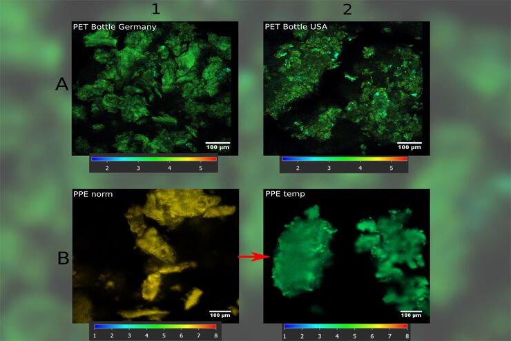
How FLIM Microscopy Helps to Detect Microplastic Pollution
The use of autofluorescence in biological samples is a widely used method to gain detailed knowledge about systems or organisms. This property is not only found in biological systems, but also…
Loading...
 - THUNDER Imager 3D Cell Culture Influenca virus – red, cilia – green, Nuclei – blue.")
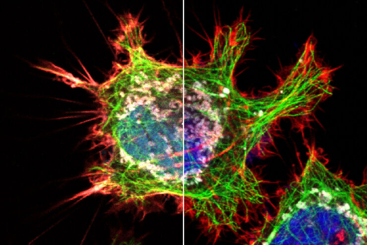
How Can Immunofluorescence Aid Virology Research?
Modern virology research has become as crucial now as ever before due to the global COVID-19 pandemic. There are many powerful technologies and assays that virologists can apply to their research into…
Loading...
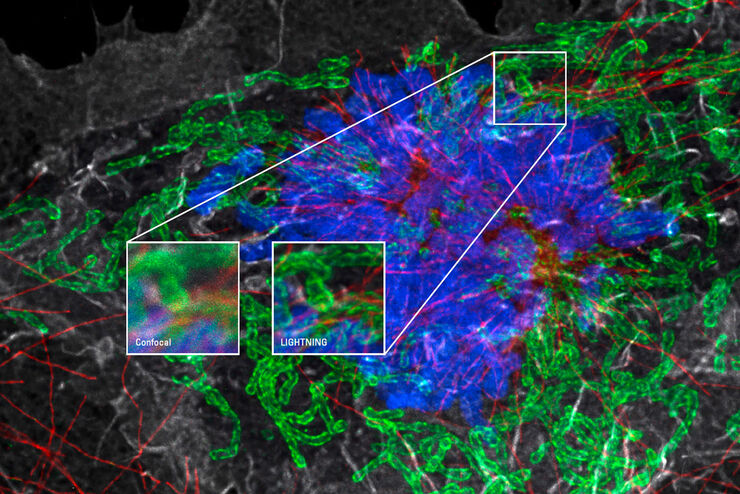
Obtain Maximum Information from your Specimen with LIGHTNING
LIGHTNING is an adaptive process for extraction of information that reveals fine structures and details, otherwise simply not visible, fully automatically. Unlike traditional technologies, that use a…
Loading...
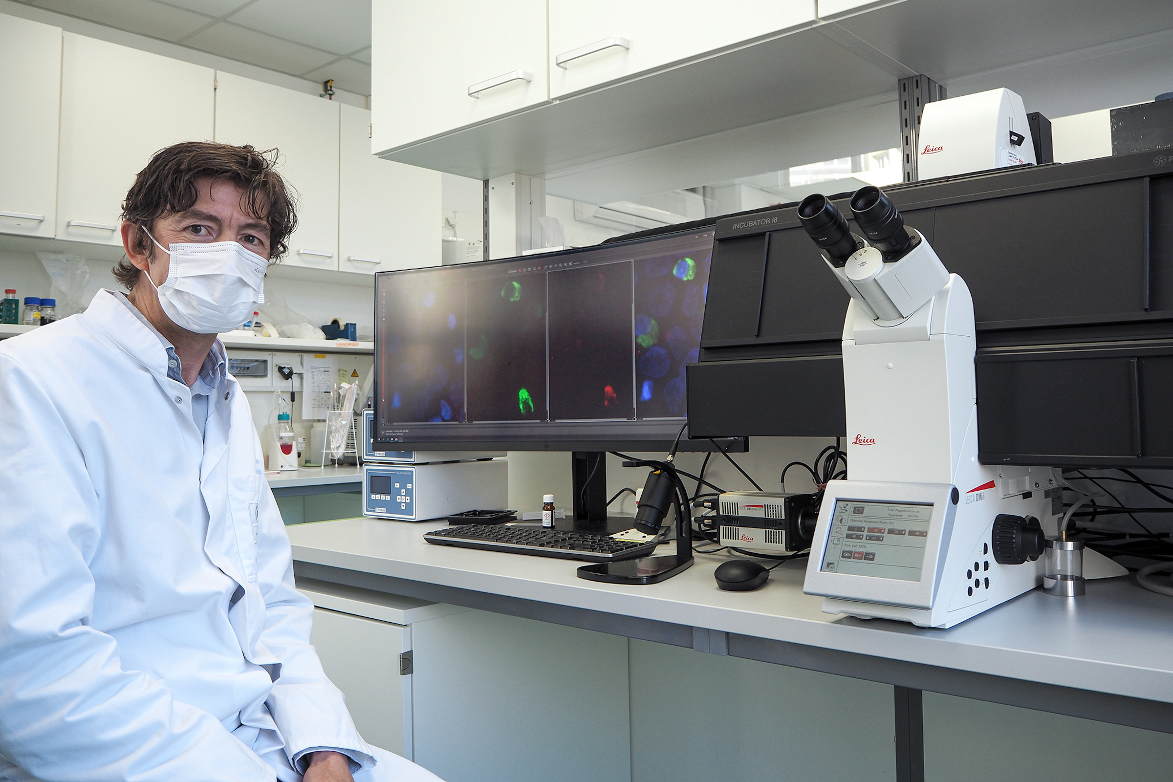
Microscopy in Virology
The coronavirus SARS-CoV-2, causing the Covid-19 disease effects our world in all aspects. Research to find immunization and treatment methods, in other words to fight this virus, gained highest…
Loading...
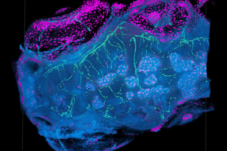
Explore Innovative Techniques to Separate Fluorophores with Overlapping Spectra
In this article we explore several strategies you can take to improve the separation of fluorophores and increase the number of fluorescent probes you can distinguish in your sample.
Loading...

STELLARIS White Light Lasers
When it comes to choosing fluorescent probes for your multi-color experiments, you shouldn’t have to compromise. Now you can advance beyond conventional excitation sources that limit your fluorophore…
Loading...
TauSense Technology Imaging Tools
Leica Microsystems’ TauSense technology is a set of imaging modes based on fluorescence lifetime. Found at the core of the STELLARIS confocal platform, it will revolutionize your imaging experiments.…
Loading...
The Power HyD Detector Family
Powerful photon counting detectors on the STELLARIS confocal platform provide improved photon counting, ultra-sensitive imaging and more color options in the NIR spectrum.
Loading...
 from the desired fluorescent signal from the cell membrane (green).")
Learn how to Remove Autofluorescence from your Confocal Images
Autofluorescence can significantly reduce what you can see in a confocal experiment. This article explores causes of autofluorescence as well as different ways to remove it, from simple media fixes to…