
Science Lab
Science Lab
The knowledge portal of Leica Microsystems offers scientific research and teaching material on the subjects of microscopy. The content is designed to support beginners, experienced practitioners and scientists alike in their everyday work and experiments. Explore interactive tutorials and application notes, discover the basics of microscopy as well as high-end technologies – become part of the Science Lab community and share your expertise!
Filter articles
Tags
Story Type
Products
Loading...
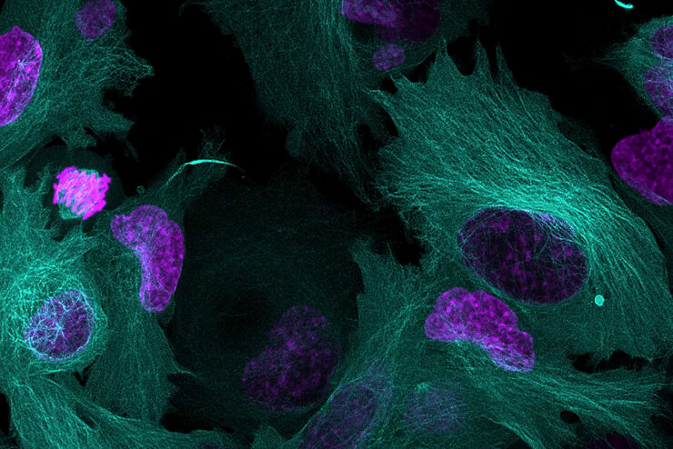
, Astrocytes (GFAP, red), Nuclei (DAPI, blue).")
Multicolor Microscopy: The Importance of Multiplexing
The term multiplexing refers to the use of multiple fluorescent dyes to examine various elements within a sample. Multiplexing allows related components and processes to be observed in parallel,…
Loading...
A New Method for Convenient and Efficient Multicolor Imaging
The technique combining hyperspectral unmixing and phasor analysis was developed to simplify the process of getting images from a sample labeled with multiple fluorophores. This aggregate method…
Loading...
Using Machine Learning in Microscopy Image Analysis
Recent exciting advances in microscopy technologies have led to exponential growth in quality and quantity of image data captured in biomedical research. However, analyzing large and increasingly…
Loading...
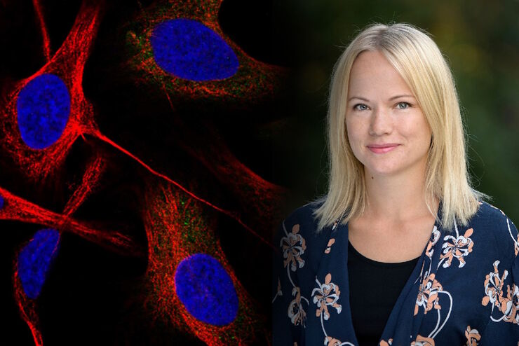
Applying AI and Machine Learning in Microscopy and Image Analysis
Prof. Emma Lundberg is a professor in cell biology proteomics at KTH Royal Institute of Technology, Sweden. She is also the director of the Cell Atlas, an integral part of the Swedish-based Human…
Loading...
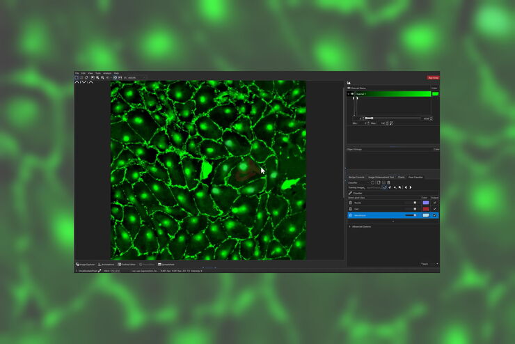
The AI-Powered Pixel Classifier
Achieving reproducible results manually requires expertise and is tedious work. But now there is a way to overcome these challenges by speeding up this analysis to extract the real value of the image…
Loading...
Considerations for Multiplex Live Cell Imaging
Simultaneous multicolor imaging for successful experiments: Live-cell imaging experiments are key to understand dynamic processes. They allow us to visually record cells in their living state, without…
Loading...

How to Improve Live Cell Imaging with Coral Life
For live-cell CLEM applications, light microscopy imaging is a critical step for identifying the right cell in the right state at the right time. In this article, Leica experts share their insights on…
Loading...
Dissecting Proteomic Heterogeneity of the Tumor Microenvironment
This lecture will highlight cutting edge applications in applying laser microdissection and microscaled quantitative proteomics and phosphoproteomics to uncover exquisite intra- and inter-tumor…
Loading...
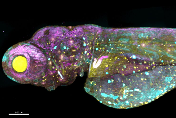
, specific neuronal markers (magenta) and cell nuclei (white), computational cleared.")
Into the Third Dimension with "Wow Effect"- Observe Cells in 3D and Real-Time
Life is fast, especially for a cell. As a rule, cells should be examined under physiological conditions which are as close as possible to their natural environment. New technologies offer tremendous…