
Science Lab
Science Lab
The knowledge portal of Leica Microsystems offers scientific research and teaching material on the subjects of microscopy. The content is designed to support beginners, experienced practitioners and scientists alike in their everyday work and experiments. Explore interactive tutorials and application notes, discover the basics of microscopy as well as high-end technologies – become part of the Science Lab community and share your expertise!
Filter articles
Tags
Story Type
Products
Loading...
 or Minor’s syndrome")
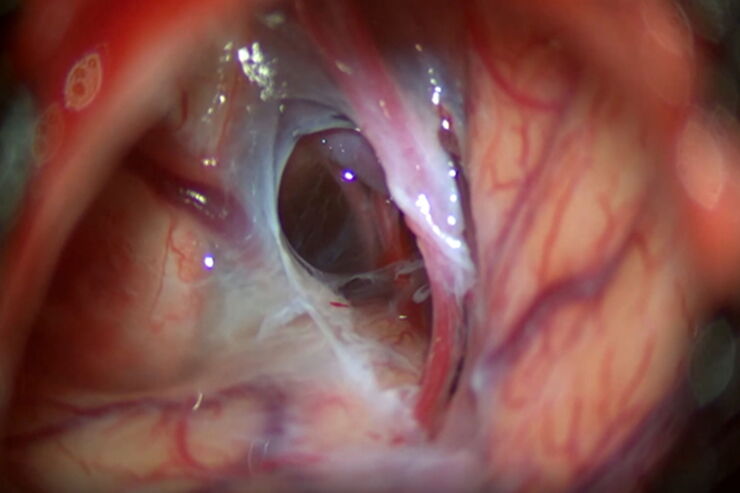
Minor’s Syndrome Surgical Intervention by Prof. Vincent Darrouzet
Minor’s disease, also called Superior Semicircular Canal Dehiscence (SSCD) or Minor’s syndrome, is a rare disorder of the inner ear that affects hearing and balance. The disease is characterized by…
Loading...
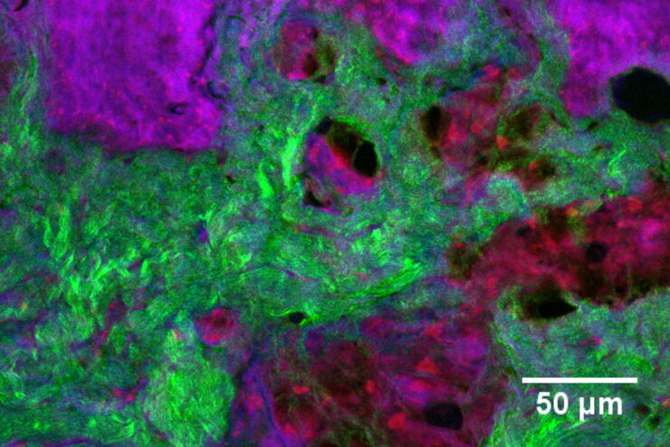
 and THUNDER (right) image of Ewing Sarcoma cells (SK-ES-1)")
Visualizing the Mitotic Spindle in Cancer Cells
This article demonstrates how this research is aided by visualizing more details of mitotic spindles in Ewing Sarcoma cells using the THUNDER Imager Tissue and Large Volume Computational Clearing…
Loading...
Formulated Product Characterization with SRS Microscopy
Creams, pastes, gels, emulsions, and tablets are ubiquitous across a wide range of manufacturing sectors from pharmaceuticals and consumer health products to agrochemicals and paint. To improve…
Loading...
Optimal Visualization in Brain Surgery
This case study “Treatment of the Galassi type III arachnoid cyst with the M530 OHX surgical microscope from Leica Microsystems” documents the procedure step by step and shows the visualization…
Loading...
Effects of Clearing Media on Tissue Transparency and Shrinkage
This study comprehensively evaluates the effects of different clearing media on tissue transparency and shrinkage by comparing freshly dissected dipteran fly brains with their cleared equivalents.…
Loading...
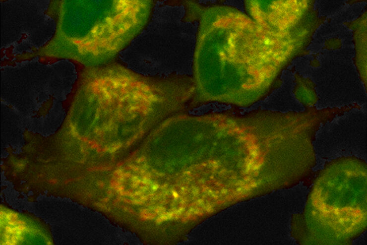
How to Quantify Changes in the Metabolic Status of Single Cells
Metabolic imaging based on fluorescence lifetime provides insights into the metabolic dynamics of cells, but its use has been limited as expertise in advanced microscopy techniques was needed.
Now,…
Loading...
, specific neuronal markers (magenta) and cell nuclei (white), computational cleared.")
Into the Third Dimension with "Wow Effect"- Observe Cells in 3D and Real-Time
Life is fast, especially for a cell. As a rule, cells should be examined under physiological conditions which are as close as possible to their natural environment. New technologies offer tremendous…
Loading...
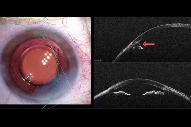
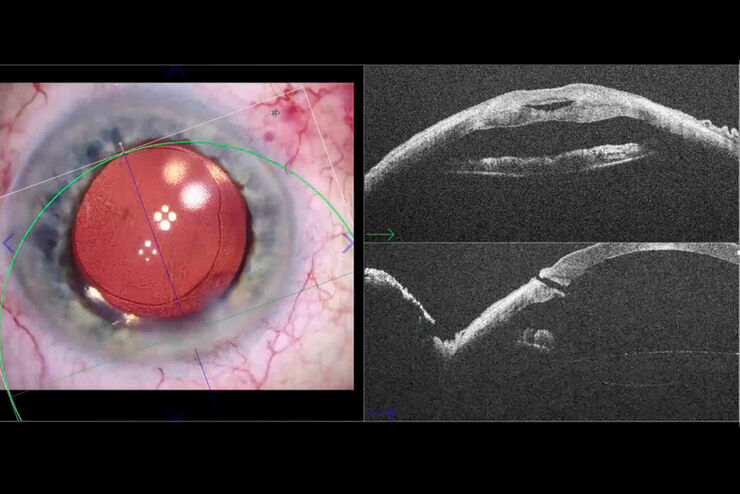
Residual Fragment Assessment with the Proveo 8 Intraoperative OCT
In the brief video presented below, Dr. Rachid Tahiri, an ophthalmic surgeon at the Centre d’Ophtalmologie in Granville, France shares his experience in using Proveo 8 intraoperative OCT in cataract…
Loading...
Cataract Incision Analysis Using EnFocus Intraoperative OCT
Ophthalmic surgery requires high levels of accuracy and precision for optimal surgical maneuvers and tissue manipulation. Surgical microscopes play an essential role in helping ophthalmic surgeons…