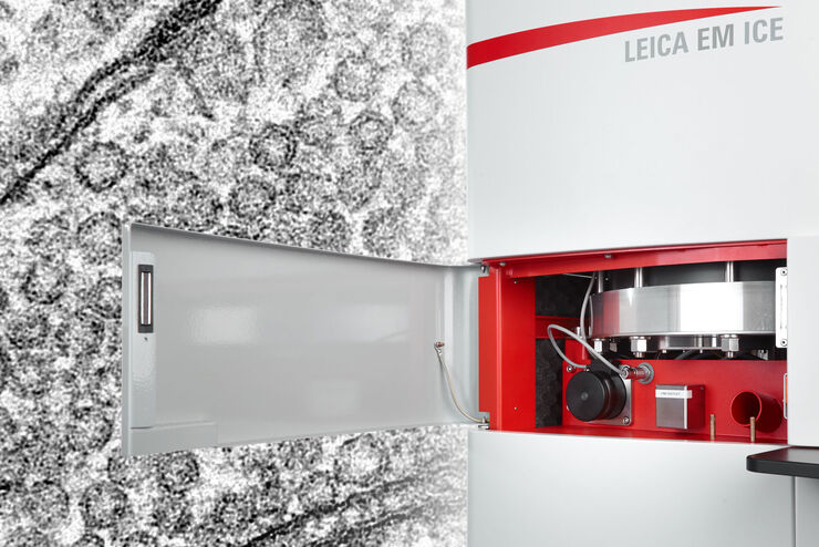
EM ICE
전자현미경 시료 전처리
제품소개
홈
Leica Microsystems
EM ICE High Pressure Freezer
빛과 전기 자극의 가능성을 가진 고압 동결
최신 기사를 읽어 보세요

How Marine Microorganism Analysis can be Improved with High-pressure Freezing
In this application example we showcase the use of EM-Sample preparation with high pressure freezing, freeze substiturion and ultramicrotomy for marine biology focusing on ultrastructural analysis of…

How to Successfully Perform Live-Cell CLEM
The Leica Nano workflow provides a streamlined live-cell CLEM solution for getting insight bout structural changes of cellular components over time. Besides the technical handling described in the…

How to Successfully Implement Coral Life
The live-cell CLEM workflow allows you to capture dynamic information related to a relevant biological process as it happens and put these observations into their ultrastructural context. The Leica…

How to Improve Live Cell Imaging with Coral Life
For live-cell CLEM applications, light microscopy imaging is a critical step for identifying the right cell in the right state at the right time. In this article, Leica experts share their insights on…

How to Keep Your Samples Under Physiological Conditions
The Coral Life workflow combines dynamic data with the best possible sample fixation by high pressure freezing. However, good sample preservation won’t help if your cells are stressed by temperature…
Capture life as it happens
With the Leica Nano Workflow, searching for the needle in the haystack is a thing of the past. Take advantage of correlative light and electron microscopy to identify directly the right cell at the…

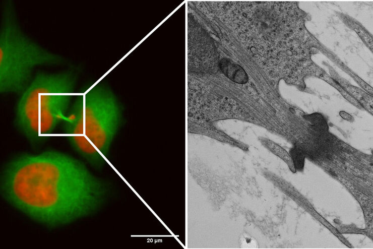
Putting Dynamic Live Cell Data into the Ultrastructural Context
With workflow Coral Life, searching for a needle in the haystack is a thing of the past. Take advantage of correlative light and electron microscopy to identify directly the right cell at the right…

Exploring the Structure and Life Cycle of Viruses
The SARS-CoV-2 outbreak started in late December 2019 and has since reached a global pandemic, leading to a worldwide battle against COVID-19. The ever-evolving electron microscopy methods offer a…

Fast, High-quality Vitrification with the EM ICE High Pressure Freezer
The EM ICE High Pressure Freezer was developed with a unique freezing principle and uses only a single pressurization and cooling liquid: liquified nitrogen (LN2). This design enables three major…

Investigating Synapses in Brain Slices with Enhanced Functional Electron Microscopy
A fundamental question of neuroscience is: what is the relationship between structural and functional properties of synapses? Over the last few decades, electrophysiology has shed light on synaptic…

High-pressure freezing: Revealing functional mechanisms of synaptic transmission
Learn more about applying optogenetic stimulation in the EM ICE and how this technology has the potential to reveal structural and functional mechanisms of synaptic transmission. Get a detailed…
Workflows and Instrumentation for Cryo-electron Microscopy
Cryo-electron microscopy is an increasingly popular modality to study the structures of macromolecular complexes and has enabled numerous new insights in cell biology. In recent years, cryo-electron…

연구 분야의 모델 유기체
모델 유기체는 연구자들이 특정한 생물학적 과정을 연구하기 위해 사용하는 종입니다. 이들은 인간과 유사한 유전적 특성을 가지고 있으며, 유전학, 발달생물학, 신경과학 같은 연구 분야에서 일반적으로 사용됩니다. 유기체 모델은 일반적으로 실험실 환경에서 쉬운 유지와 번식, 짧은 세대 주기 또는 특정 형질이나 질병을 연구하기 위한 돌연변이 생성 능력 때문에…

바이러스학
연구의 관심 분야가 바이러스 감염과 질병에 집중되어 있습니까? 라이카마이크로시스템즈의 이미징 및 샘플 준비 솔루션을 통해 바이러스학에 관한 통찰력을 얻는 방법을 알아보세요.

Expert Knowledge on High Pressure Freezing and Freeze Fracturing in the Cryo SEM Workflow
Get an insight in the working methods of the laboratory and learn about the advantages of Cryo SEM investigation in EM Sample Preparation. Find out how high pressure freezing, freeze fracturing and…
Bridging Structure and Dynamics at the Nanoscale through Optogenetics and Electrical Stimulation
Nanoscale ultrastructural information is typically obtained by means of static imaging of a fixed and processed specimen. However, this is only a snapshot of one moment within a dynamic system in…
Expanding the Limits of Electron Microscopy Sample Preparation
Capturing the intricate changes in fine structure or in cell dynamics with conventional cryo solutions can be challenging sometimes. Leica Microsystems has developed a new cryo platform, the Leica EM…
Interview with Dr. Shigeki Watanabe on Research in Synaptic Membrane Dynamics
Dr. Shigeki Watanabe, principle investigator of the department of Cell Biology at the Johns Hopkins University School of Medicine in Baltimore, held a workshop in Zürich, Switzerland on methods to…
Fields of Application
상관관계적 광학 현미경과 전자현미경(CLEM)
라이카마이크로시스템즈 Coral 작업 흐름은 사용자가 형광 현미경과 전자현미경 (CLEM) 데이터의 상관관계를 분석하는 데 도움이 됩니다.
