
Material and Methods for plankton analysis
Dinoflagellates were collected by towing a net (5 or 10 µm mesh size) for 10 minutes at slow speed in surface waters of the Villefranche-sur-Mer bay (France). Upon collection onboard, samples were then filtered through serial sieves (Retsch) to collect cells below 40 µm in size. In the laboratory, this size fraction was 1) concentrated by filtration on a 1.2 mixed cellulose ester membrane (Merck) using manual pumping; 2) resuspended in a final volume of about 4 ml, and 3) centrifuged for 5 minutes at 1000g. The supernatant was removed and ca 1.2 µl of the pellet was loaded in a gold copper type A carrier (Leica; 200 µm deep and 3 mm wide) precoated with hexadecene (Merck). The sample was covered by the flat side of an aluminium type B carrier (Leica) precoated with hexadecene (Merck) as well, and high-pressure frozen using the Leica EM ICE.

 and B carrier (3 mm wide, flat side was used).")
Cryofixed samples underwent freeze-substitution (EM AFS2, Leica) using the following program and solutions: 60h at -90°C in 2% osmium tetroxide in acetone, heating rate of 2°C/h for 15h to -60°C, 10h at -60°C, heating rate of 2°C/h for 15h to -30°C, 10h at -30°C, maximum heating rate to 0°C in 1 minute, 1h at 0°C, maximum cooling rate to -30°C in 1 minute, 5 washes at -30°C using pure acetone.
Cells were then gradually infiltrated with EPON hard. The resin (without accelerator)/acetone (v/v) series used were 25% for 2h starting at -30°C and reaching -10°C, 50% for 2h starting at -10°C and reaching +10°C with a heating rate of 5°C/h and 75% for 2h starting at +10°C and reaching +20°C with a heating rate of 10°C/h. Samples were then infiltrated in 100% resin without accelerator successively for 12 and 48 hours.
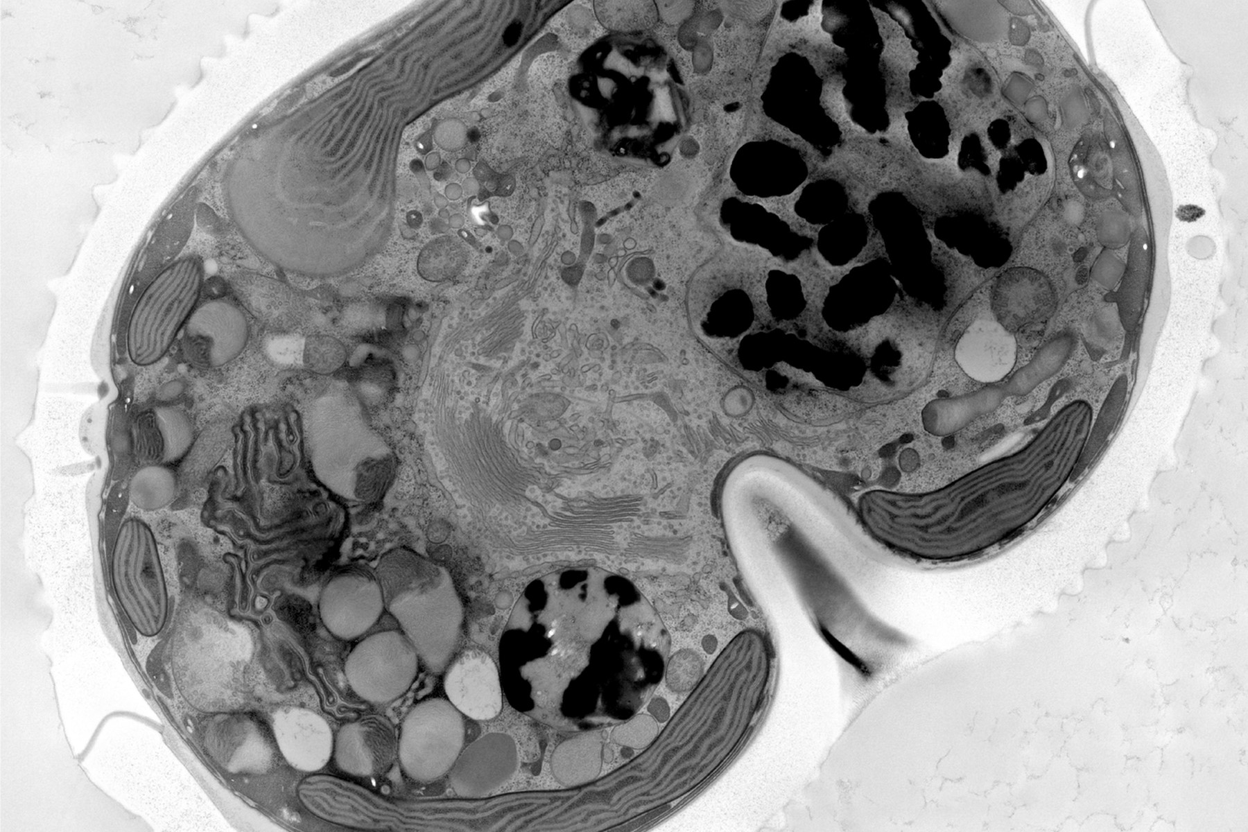
Infiltration with 100% resin containing accelerator was then performed for 2 times 3h and one time overnight before polymerization at 60°C for 48 hours. 70nm thin sections were obtained using an ultramicrotome (Leica EM UC7) with an ultra-diamond knife (Diatome). Thin sections were then post stained using successively 1% uranyl acetate in water for 20 minutes and lead citrate for 3 minutes. Tile montages were acquired through SerialEM on the JEOL JEM 2100plus using 120Kv and 8000X magnification. Montages were processed using Imod and Fiji. High pressure frozen samples were processed in the Electron Core Microscopy Facility (EMCF) at EMBL Heidelberg.
Results


 labeled with membrane-permeable calcein, high-pressure frozen in salt water using EM ICE.")
