
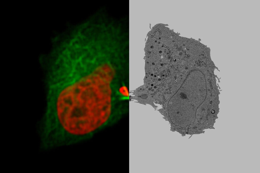
Dynamic processes in their ultrastructural context
Now there is no need to guess about the underlying mechanisms when analyzing a wide range of biological events, like intracellular trafficking, nuclear envelope formation, or mitotic spindle assembly. The workflow Coral Life is a proven solution, combining live-cell correlative light and electron microscopy (CLEM) with time-specific fixation by high-pressure freezing, which enables the investigation of dynamic events with nanometer resolution. A massive surplus of context data can be obtained by the combination of dynamic data, specifically targeted entities by fluorescent live-cell imaging, and precisely timed EM ultrastructural analysis.
The right balance of patience, timing, and speed
Many electron microscope workflows start with the fixation of cells, followed by sample preparation and EM imaging. However, cells displaying interesting behavior tend to be rare. Making things even more complicated is the fact that the cellular behavior or processes of interest might be very transient. Finding the “right cell” is often very time-consuming and tedious work which seems like “looking for a needle in a haystack”.
The Live-cell CLEM workflow allows you to capture dynamic information of a relevant biological process when it happens and enables you to put these observations into their ultrastructural context. With Coral Life, you can rest assured that you obtain the results you are looking for. Find the right cell in the right state at the right time with the light microscope, observe it, and retrieve it rapidly and reliably once the sample is loaded in the electron microscope. Thanks to the dedicated SampLink chamber, you enjoy peace of mind while looking for the cell of interest with the light microscope and quickly “snap-freeze” it precisely at the correct moment.
Be relaxed
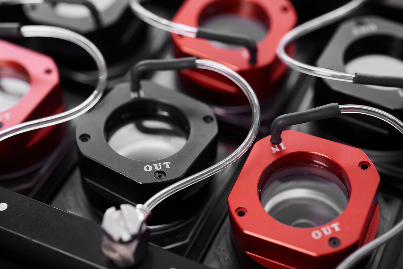
Due to the open chamber design and continuous gas exchange the SampLink chambers keep live cells healthy, giving you enough time to select and image a region of interest until you find cells in the exact state you are looking for. There is no pressure to make decisions quickly before your cells suffer a decline in health.
Be quick and precise
Within 5 seconds after you have identified your time point of interest, the SampLink chamber is transferred to the EM ICE and the cells are fixed by high pressure freezing for further downstream analysis.
An imaging window for dynamic processes
A transfer time of just 5 seconds opens a window to a wide range of dynamic processes which occur in living cells. With some experience, you can even refine the exact point in time of the processes which you want to capture in the prepared specimens. Whether you are interested in vesicle transport and sorting, filopodial dynamics, endocytosis, cell migration, cytokinesis, or a plethora of other biological processes, Coral Life gives you complementary dynamic and ultrastructural information.
The generous time window before the transfer ensures that you to have enough room for sample identification and imaging. Without the pressure to make a fast decision, typical of other live-cell CLEM systems where often there are only a few minutes to choose a region, you can make the selection more calmly, the success rate increases, and the results are of better quality and more reproducible.

A streamlined workflow for reproducible results
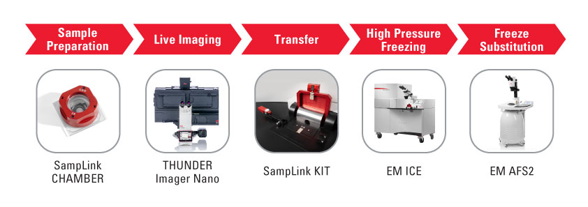
Sample Preparation
Keep your samples under optimal conditions
With the SampLink chamber, culturing, transport, and imaging of cells on sapphire is possible without any physiological interruption, even when transporting cells between the lab and microscopy facility.
- Permits long term imaging under physiological conditions thanks to the open sample container with continuous gas exchange
- Gas and temperature control during cell cultivation and transport with Oxygenie’s incubator system

Live Imaging
Optimized image resolution
For optimal resolution during live-cell imaging, the THUNDER Imager Nano is equipped with a specific sapphire-adapted immersion objective. It enables high resolution and simplifies live-cell imaging, as one does not need to use dip-in objectives which increase the risk of contaminating the cells. SampLink chambers can be loaded directly into the THUNDER Imager Nano.
- Fast 3D live-cell imaging for a more accurate study of physiology and blur removal for better target identification with THUNDER
- Stable physiological conditions of the sample due to the connection to the Oxygenie system and temperature control via the microscope
- Optimized image resolution with the equipped 40x / 1.1 NA sapphire-corrected water-immersion objective

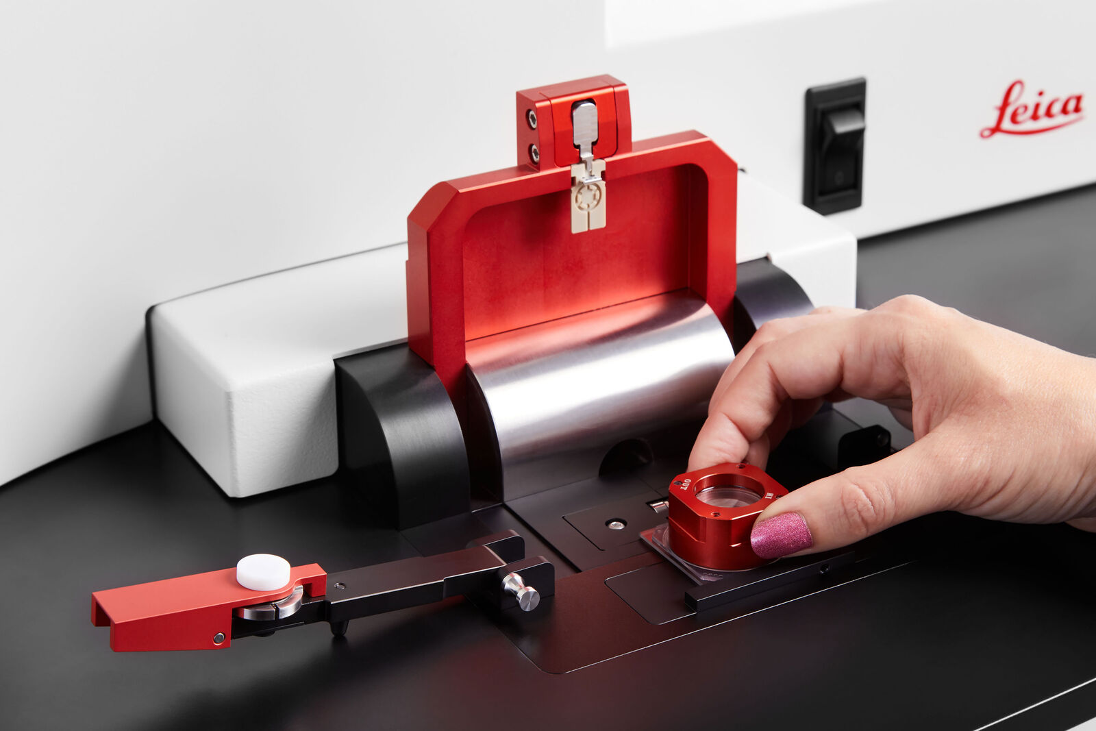
Transfer
Time is key
To minimize the time between live-cell imaging and freezing, the workflow Coral Life comes with an intuitive transfer system that allows users to start the freezing directly after imaging, while keeping the sample under physiological control.
- Capture transient events with a transfer that takes less than 5 seconds
- Benefit from an all-in-one package with complementary consumables
- Adapt easily and quickly to the EM ICE with the slide-2-release mechanism for the middle plate

High Pressure Freezing
Optimal sample fixation
With the EM ICE, the imaged samples are ultimately cryo-immobilized. It is the optimal system for combining classic high-pressure freezing with new workflow solutions. Not only can it be retrofitted to Coral Life, but it can also be combined with light stimulation experiments.
- Full vitrification of adherent cells
- Live-cell CLEM can be combined with optical stimulation experiments thanks to the optional upgrade for light and electrical stimulation
- Optimal results with the market-leading solution

Freeze Substitution
Downstream workflows
Benefit from our established freeze substitution downstream workflow using the EM AFS2, EM TRIM2, EM UC7, or EM AC20 to prepare your samples for subsequent TEM imaging. Coral Life is also suitable for SEM tomography analysis. Use the ARTOS 3D to prepare samples on wafers.

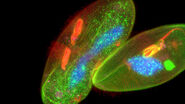
 image of a cross section of C. elegans (roundworm). Courtesy of T. Müller-Reichert, MPI-CBG, Dresden, Germany and K. McDonald, University of California, Berkeley, USA.")


