Leica LMD6 & LMD7
정립 현미경
광학 현미경
제품소개
홈
Leica Microsystems
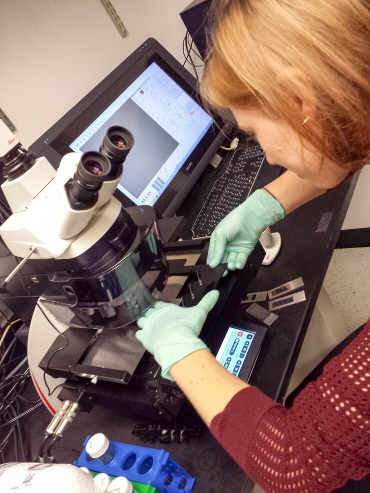
Leica LMD6 & LMD7 레이저 미세절단 현미경 시스템
완벽한 해부
최신 기사를 읽어 보세요

Transforming Research with Spatial Proteomics Workflows
Spatial Proteomics, Nature Methods 2024 Method of the Year, is driving research advancements in cancer, immunology, and beyond. By combining positional data with high throughput imaging of proteins in…

Deep Visual Proteomics Provides Precise Spatial Proteomic Information
Despite the availability of imaging methods and mass spectroscopy for spatial proteomics, a key challenge that remains is correlating images with single-cell resolution to protein-abundance…

A Guide to Spatial Biology
What is spatial biology, and how can researchers leverage its tools to meet the growing demands of biological questions in the post-omics era? This article provides a brief overview of spatial biology…

An Introduction to Laser Microdissection
The heterogeneity of histological and biological specimens often requires isolation of specific single cells or cell groups from surrounding tissue before molecular biology analysis can be carried…
 microdissected with a 10x objective (upper right). Inspection of the collection device (lower right).")
Molecular Biology Analysis facilitated with Laser Microdissection (LMD)
Extracting biomolecules, proteins, nucleic acids, lipids, and chromosomes, as well as extracting and manipulating cells and tissues with laser microdissection (LMD) enables insights to be gained into…
.")
Spatial Metabolomics: Exploring Tumor Complexity and Therapeutic Insights
In cancer research, it is vital to understand the interaction between tumor cells and their microenvironment, as the tumor microenvironment influences tumor progression significantly. Spatial…

Lipidomics Analysis of Sparse Cells based on Laser Microdissection
Delve into cellular intricacies with high-coverage targeted lipidomics analysis of sparse cells. This advanced method, integrating Laser Microdissection (LMD) and Liquid Chromatography-Mass…
.")
Neuron Isolation in Spatial Context with Laser Microdissection (LMD)
After Alzheimer’s disease, Parkinson’s is the second most common progressive neurodegenerative disease. Before the first symptoms manifest, up to 70% of dopamine-releasing neurons in the mid-brain…

How did Laser Microdissection enable Pioneering Neuroscience Research?
Dr. Marta Paterlini, a Senior Scientist at the Karolinska Institute, shares her experience of using laser microdissection (LMD) in groundbreaking research into adult human neurogenesis and offers…

Laser Microdissection Protocols for Tissue and Cell Isolation - Download free eBook
In this Bio-protocol Selections, we present a collection of open-access, detailed methods papers using LCM to purify and isolate tissues and cells from plants, mouse embryos, cancer cells, neurons,…

Studying Virus Replication with Fluorescence Microscopy
The results from research on SARS-CoV-2 virus replication kinetics, adaption capabilities, and cytopathology in Vero E6 cells, done with the help of fluorescence microscopy, are described in this…

Exploring Subcellular Spatial Phenotypes with SPARCS
Discover spatially resolved CRISPR screening (SPARCS), a platform for microscopy-based genetic screening for spatial subcellular phenotypes at the human genome scale.

Spatial Biology: Learning the Landscape
Spatial Biology: Understanding the organization and interaction of molecules, cells, and tissues in their native spatial context

How is Microscopy Used in Spatial Biology? A Microscopy Guide
Different spatial biology methods in microscopy, such as multiplex imaging, are helping to better understand tissue landscapes. Learn more in this microscopy guide.

Dissecting Proteomic Heterogeneity of the Tumor Microenvironment
This lecture will highlight cutting edge applications in applying laser microdissection and microscaled quantitative proteomics and phosphoproteomics to uncover exquisite intra- and inter-tumor…

20 Years of Leica Laser Microdissection
Phenotype-genotype correlations are key for insight. From Eye to Insight is therefore fitting perfectly to Leica Microsystems and in particular to laser microdissection. Laser Microdissection, also…

Consumables for Laser Microdissection
There are many different types of consumables for laser microdissection (LMD) systems. They cover a wide range of applications from basic to highly specialized, enabling scientists to choose their own…

바이러스학
연구의 관심 분야가 바이러스 감염과 질병에 집중되어 있습니까? 라이카마이크로시스템즈의 이미징 및 샘플 준비 솔루션을 통해 바이러스학에 관한 통찰력을 얻는 방법을 알아보세요.
Workflows & Protocols: How to Use a Leica Laser Microdissection System and Qiagen Kits for Successful RNA Analysis
Laser Microdissection (LMD) allows isolating individual cells or chromosomes and is a well established technique for sample preparation prior downstream analysis of the nucleic acid content via PCR or…

Forensic Detection of Sperm from Sexual Assault Evidence
The impact of modern scientific methods on the analysis of crime scene evidence has dramatically changed many forensic sub-specialties. Arguably one of the most dramatic examples is the impact of…
Fields of Application
법의학
과학수사에서 현미경 및 이미징 장비는 증거를 조사 할때 정확성을 위하여 결과의 정밀도, 품질, 정확성 및 재현성을 제공해야합니다. 라이카사는 일반 실험장비에서 완전 자동화 시스템까지 광범위한 현미경 시스템을 사용하여 정량, 분석 및 문서화 작업을 지원 해줍니다.
