
The principle of laser microdissection (LMD)
To perform microdissection, an upright or inverted microscope is coupled to a laser. Selected areas of a specimen or even individual cells are then excised by moving the focused laser beam along their contours. This procedure guarantees gentle specimen handling – in particular, it does not transfer any heat to the specimen – and provides the exact material that is needed for analysis.
The dissectate is then transported to a collection device in a number of different ways. Depending on the system used, dissected tissue either falls under the force of gravity into a reaction vessel (Leica LMD system, refer to figure 1), is catapulted against the force of gravity into a reaction vessel, or removed indirectly together with a membrane covering it.
What is laser microdissection used for?
Laser microdissection is an established method for a large number of applications, mainly those of molecular biology, particularly nucleic acid research, neurosciences, developmental biology, cancer research, forensics, proteomics, plant research, cutting cell cultures, and single cell isolation [1]. Laser microdissection is even used in climate research, especially for examining the annual rings of trees.
 Defining the region of interest; Step 2) Steering the laser beam with the optics along the cut line; and Step 3) Collecting the specimen with gravity.")
Methods and technology of non-contact laser microdissection
Laser microdissection offers a precise and contamination-free solution for the isolation and selection of single cells or tissue. The tissue samples can be embedded, sectioned, and stained according to conventional methods of preparation. Paraffin sections, frozen sections, smear preparations, chromosome specimens, and cell cultures are all suitable for laser microdissection. The area selected for dissection is drawn on the PC screen and automatically separated from the surrounding tissue with a laser beam. Fluorescence-labelled specimens can also be dissected using special filter cubes which transmit the full spectrum of laser light. The dissectate is then immediately transported to a collection device (in different ways, depending on the manufacturer of the system) for further examination.
The force of gravity is the method of choice for collection with the Leica LMD system. Here the sample is fixed and the laser beam is moved over it. This approach allows easy viewing of the sample and collecting of the dissectate which falls into a receptable device.
This approach allows the use of standard lab material for collection, ranging from single PCR tubes to well plates. Direct collection into well plates is critical for higher throughput, since pipetting steps from single PCR collection tubes into well-plates became obsolete. One of the workflows which benefit from this time-saving step is Deep Visual Proteomics (DVP) where the abundance of proteins is correlated with their spatial location at the single cell level [2].
The precision of the laser-cutting process is optically coupled to the chosen magnification. A higher magnification automatically results in a finer step width, as the laser beam and its movement are reduced by the same degree as the field of view. No other work steps are required in this case. Not only single cells, but also larger areas can be excised in a single pass. When transferring the dissectate to a collection device, there is no risk of contact or contamination.

As seen in figure 2, the complete process required for LMS and molecular biology analysis has 5 steps:
- Specimen Preparation: Selection and prepare biological specimens (Histological specimens, Living cells and cell cultures, Chromosome spreads, Smears, Cytospins, Plant material, Sperm and other forensic preparations)
- Staining: Visualize regions of interest (ROI)
- Brightfield: HE (hematoxylin-eosin), Cresyl violet, Toluidin blue, Thionin, Immunohistochemistry
- Fluorescence: Secondary antibodies, Acridine orange, FISH
- Laser Microdissection: Selectively dissect ROIs
- Contact-free, Contamination-free, Any size from cell compartments of a few µm2 to several mm2, Any shape
- Extraction: Extract and prepare important molecules
- DNA, RNA, Proteins, Metabolites, Biomarkers
- Analysis: Obtain reproducible and specific results
- PCR, Quantitative real-time PCR, Microarrays, Expression profiling, Genetic fingerprinting, LOH, FISH, LC-MS/MS, 2-D PAGE, SELDI, MALDI
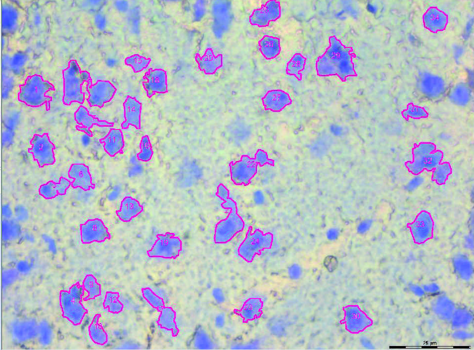
Example of preparing samples from tissue with LMD
The preparation of dissectates from a 12-μm thick brain tissue section using LMD is shown in figure 3. The brain section was stained with Toluidin blue before microdissection. The images shown were taken with a Leica microscope using a 63x objective. The detection of shapes was done by the ADM (Auto Detection Module). The selected shapes are within the chosen detection criteria.
 Image of a 12-μm thick brain section before laser microdissection and B) the same 12- μm thick brain section after dissection.")

 and astrocytes (green) in a cortical spheroid derived from human induced pluripotent stem cells.")

