THUNDER Imager Tissue
THUNDER Imaging Systems
제품소개
홈
Leica Microsystems
THUNDER Imager Tissue
실시간 3차원 바이오이미지 디코딩*
최신 기사를 읽어 보세요

제브라피시 연구
선별, 분류, 조작 및 이미징 중 최상의 결과를 얻으려면 세부 사항과 구조를 관찰해 다음 연구 단계를 위한 올바른 결정을 내려야 합니다.
탁월한 광학 성능과 우수한 해상도로 유명한 Leica 실체 현미경과 투과광 베이스는 전 세계 연구자들이 선택하는 제품입니다.

Improving Zebrafish-Embryo Screening with Fast, High-Contrast Imaging
Discover from this article how screening of transgenic zebrafish embryos is boosted with high-speed, high-contrast imaging using the DM6 B microscope, ensuring accurate targeting for developmental…
 acquired using THUNDER Imager Live Cell. Image courtesy of Janina Kaspar and Irene Santisteban, Schäfer Lab, TUM.")
Imaging Organoid Models to Investigate Brain Health
Imaging human brain organoid models to study the phenotypes of specialized brain cells called microglia, and the potential applications of these organoid models in health and disease.
. Image courtesy of Prof. Hui Guo, School of Life Sciences, Central South University, China")
How Microscopy Helps the Study of Mechanoceptive and Synaptic Pathways
In this podcast, Dr Langenhan explains how microscopy helps his team to study mechanoceptive and synaptic pathways, their challenges, and how they overcome them.

What are the Challenges in Neuroscience Microscopy?
eBook outlining the visualization of the nervous system using different types of microscopy techniques and methods to address questions in neuroscience.

Going Beyond Deconvolution
Widefield fluorescence microscopy is often used to visualize structures in life science specimens and obtain useful information. With the use of fluorescent proteins or dyes, discrete specimen…

Find Relevant Specimen Details from Overviews
Switch from searching image by image to seeing the full overview of samples quickly and identifying the important specimen details instantly with confocal microscopy. Use that knowledge to set up…

Accurately Analyze Fluorescent Widefield Images
The specificity of fluorescence microscopy allows researchers to accurately observe and analyze biological processes and structures quickly and easily, even when using thick or large samples. However,…

High-resolution 3D Imaging to Investigate Tissue Ageing
Award-winning researcher Dr. Anjali Kusumbe demonstrates age-related changes in vascular microenvironments through single-cell resolution 3D imaging of young and aged organs.

Optimizing THUNDER Platform for High-Content Slide Scanning
With rising demand for full-tissue imaging and the need for FL signal quantitation in diverse biological specimens, the limits on HC imaging technology are tested, while user trainability and…

Physiology Image Gallery
Physiology is about the processes and functions within a living organism. Research in physiology focuses on the activities and functions of an organism’s organs, tissues, or cells, including the…

암시야 현미경
암시야 대비법은 재료 시료의 불균일한 특징부 또는 생물학적 표본의 구조로부터 광의 회절 또는 산란을 이용합니다.

Developmental Biology Image Gallery
Developmental biology explores the development of complex organisms from the embryo to adulthood to understand in detail the origins of disease. This category of the gallery shows images about…

The Power of Pairing Adaptive Deconvolution with Computational Clearing
Learn how deconvolution allows you to overcome losses in image resolution and contrast in widefield fluorescence microscopy due to the wave nature of light and the diffraction of light by optical…
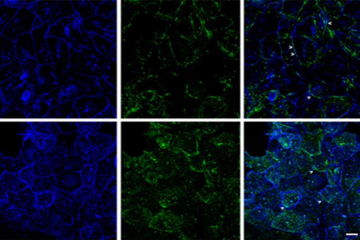
Improvement of Imaging Techniques to Understand Organelle Membrane Cell Dynamics
Understanding cell functions in normal and tumorous tissue is a key factor in advancing research of potential treatment strategies and understanding why some treatments might fail. Single-cell…

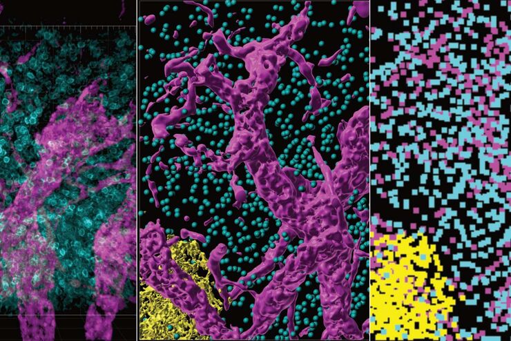
Image Gallery: THUNDER Imager
To help you answer important scientific questions, THUNDER Imagers eliminate the out-of-focus blur that clouds the view of thick samples when using camera-based fluorescence microscopes. They achieve…
From Organs to Tissues to Cells: Analyzing 3D Specimens with Widefield Microscopy
Obtaining high-quality data and images from thick 3D samples is challenging using traditional widefield microscopy because of the contribution of out-of-focus light. In this webinar, Falco Krüger…

An Introduction to Computational Clearing
Many software packages include background subtraction algorithms to enhance the contrast of features in the image by reducing background noise. The most common methods used to remove background noise…
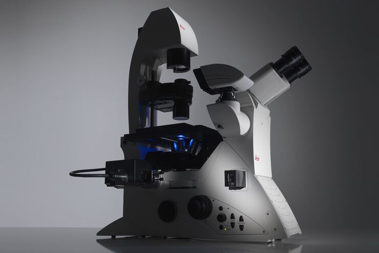
Factors to Consider When Selecting a Research Microscope
An optical microscope is often one of the central devices in a life-science research lab. It can be used for various applications which shed light on many scientific questions. Thereby the…

Computational Clearing - Enhance 3D Specimen Imaging
This webinar is designed to clarify crucial specifications that contribute to THUNDER Imagers' transformative visualization of 3D samples and improvements within a researcher's imaging-related…

암 연구
암은 성장 통제에 결함이 있는 세포에 의해 발생하는 복잡하고 이질적인 질병입니다. 하나 또는 한 그룹의 세포에서 일어나는 유전적 또는 후생적 변화가 정상적인 기능을 방해하고, 자율적이고 통제되지 않는 세포 성장과 증식을 초래합니다

THUNDER Imagers: High Performance, Versatility and Ease-of-Use for your Everyday Imaging Workflows
This webinar will showcase the versatility and performance of THUNDER Imagers in many different life science applications: from counting nuclei in retina sections and RNA molecules in cancer tissue…
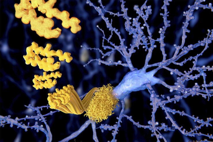
Alzheimer Plaques: fast Visualization in Thick Sections
More than 60% of all diagnosed cases of dementia are attributed to Alzheimer’s disease. Typical of this disease are histological alterations in the brain tissue. So far, there is no cure for this…
 and YOYO 1 iodide (Nucleus).")
Real Time Images of 3D Specimens with Sharp Contrast Free of Haze
THUNDER Imagers deliver in real time images of 3D specimens with sharp contrast, free of the haze or out-of-focus blur typical of widefield systems. They can even image clearly places deep inside a…
