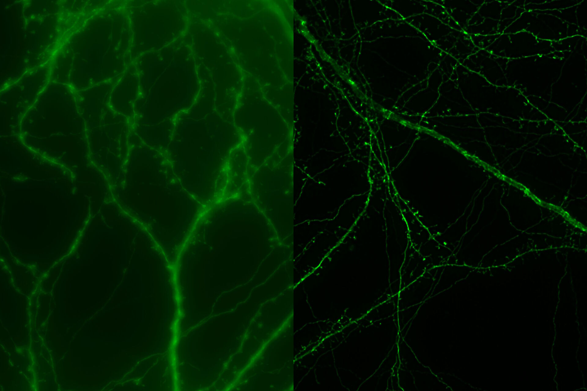
. Image courtesy of Prof. Hui Guo, School of Life Sciences, Central South University, China")
About Tobias Langenhan and his group’s work
Tobi Langenhan specializes in synaptic communication and signalling pathways that are relayed through specific types of surface receptor molecules. In his group’s work, microscopy plays an important role in helping to understand where proteins such as adhesion G-protein coupled receptors (GPCRs), or synaptic molecules that execute neurotransmission, are located within cells, and how they move and interact with other molecules. This information is vital in understanding the workings of different molecules and what they do within individual cell types, within an organ and within the context of an entire body.
Much of the microscopy work done by the group involves live cell imaging, often using model organisms, although they also use histological sections of organs of interest – specifically from the fruit fly. Different labelling strategies are employed within the lab, from immunohistochemical approaches in conjunction with molecular labelling techniques to more recent approaches such as labelling cell-surface proteins using click chemistry.
The podcast
In this podcast, Tobi discusses these labelling strategies and microscopy approaches in more detail, talking about some of the challenging questions they are trying to answer. He explains how they could obtain highly resolved images of intact model organisms using THUNDER Imaging technology, and what the future holds for his research.


 at 2 weeks. Image acquired using Mica.")