
is mobile? false
Aplicações
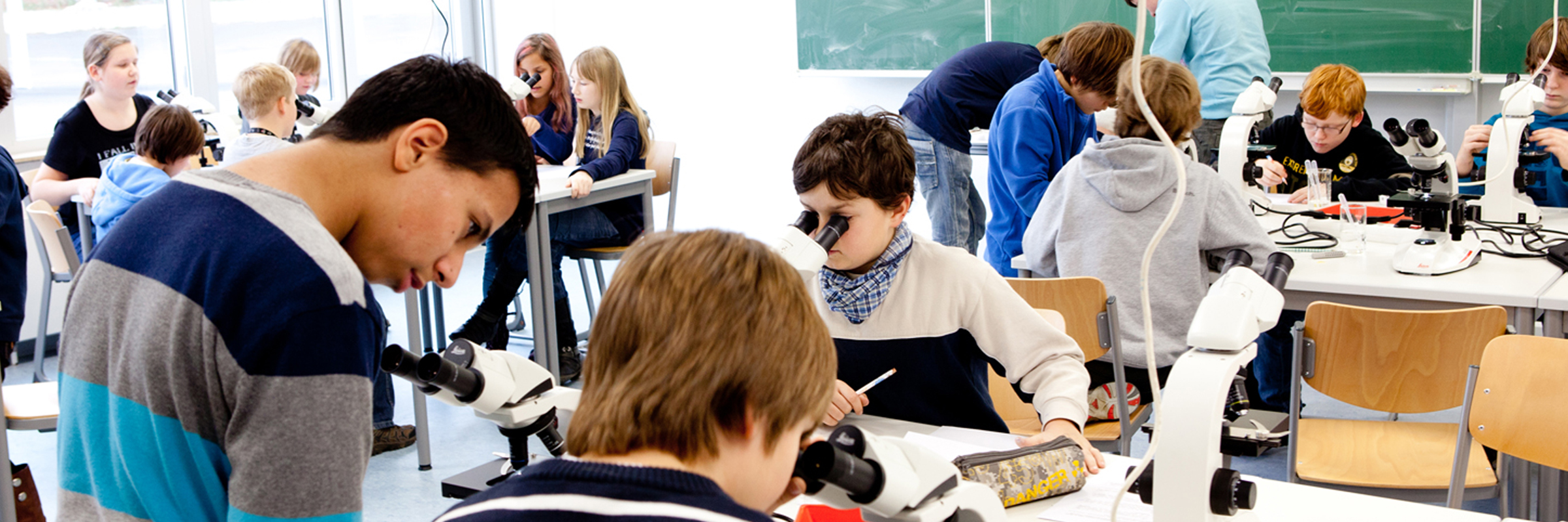
Aplicações
A microscopia é importante em muitas áreas de trabalho. As aplicações são múltiplas. Os microscópios e os instrumentos científicos que oferecemos são as ferramentas que o ajudam a atingir seus objetivos. Se você precisa lidar com tarefas repetitivas, de alto rendimento ou com questões de pesquisa muito específicas, certamente poderemos oferecer-lhe uma solução de imagem completa para sua finalidade. Nossas equipes de vendas e especialistas de aplicações com seus conhecimentos aprofundados podem ajudá-lo a encontrar um sistema de imagem ideal ou um fluxo de trabalho de preparação de amostras de microscopia eletrônica.
Selecione sua area de aplicação Show subnavigation
Aplicações
Find the solution that fits
Leica Science Lab Articles Show subnavigation
Leia os nossos artigos mais recentes
O portal de conhecimentos da Leica Microsystems oferece materiais para a pesquisa científica e ensino sobre assuntos na área de microscopia. O conteúdo foi criado para dar apoio a iniciantes, praticantes experientes e também a cientistas nos seus trabalhos e experiências diárias.
How Fluorescence Guides Sectioning of Resin-embedded EM Samples
Electron microscopes, including transmission electron microscopes (TEM) and scanning electron microscopes (SEM), are widely utilized to gain detailed structural information about biological samples or non-living materials. Ultramicrotomy is the preferred technique for producing ultrathin sections, less than 100 nm thick for TEM/SEM analysis. During sample preparation small sample pieces are embedded in epoxy or acrylic resin, excess resin is trimmed away, and the specimen is sliced into ultrathin sections (50 nm - 100 nm) using a glass or diamond knife.
Coherent Raman Scattering Microscopy Publication List
CRS (Coherent Raman Scattering) microscopy is an umbrella term for label-free methods that image biological structures by exploiting the characteristic, intrinsic vibrational contrast of their molecules. The two most important CRS techniques are Coherent Anti-Stokes Raman Scattering (CARS) and Stimulated Raman Scattering (SRS). The biochemical image contrast of CRS is in many ways complementary to the molecular contrast obtained in fluorescence microscopy. A second crucial advantage of these methods is that they preserve the specimen/sample in a near pristine state. This reference list presents current and basic papers on CRS microscopy.
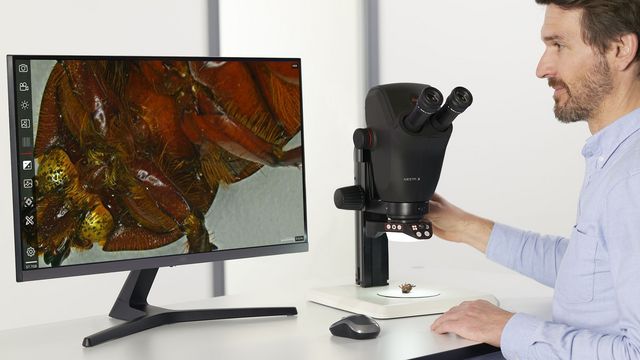
Microscópios de dissecação
Você pode passar muitas horas olhando através de oculares de um microscópio de dissecação sempre que for necessário realizar dissecações. A Leica Microsystems permite que você escolha entre uma série diversa de microscópios e uma ampla gama de peças e acessórios de microscópios de dissecação, para que você possa encontrar a solução de microscopia certa para atender às suas necessidades.
Get to Insights Faster and Easier with AI Image Analysis Tools
Discover how Aivia helps scientists streamline image analysis with fast setup, accurate AI detection, and easy batch processing.

Unlocking the Secrets of Organoid Models in Biomedical Research
Get ready to delve deeper into the world of organoids and 3D models, which are essential tools for advancing our understanding of human health. Navigating these complex structures and obtaining clear images for analysis can be challenging. In this event, researchers from The University of Oxford and University College London will join us to show how the THUNDER Imager Cell Spinning Disk can provide more convincing, high-quality data for deeper insights into a variety of models.

A Guide to Polarized Light Microscopy
Polarized light microscopy (POL) enhances contrast in birefringent materials and is used in geology, biology, and materials science to study minerals, crystals, fibers, and plant cell walls.
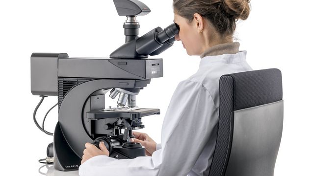
Factors to Consider when Selecting Clinical Microscopes
What matters if you would like to purchase a clinical microscope? Learn how to arrive at the best buying decision from our Science Lab Article.
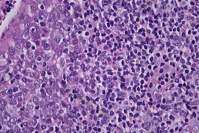
Clinical Microscopy: Considerations on Camera Selection
The need for images in pathology laboratories has significantly increased over the past few years, be it in histopathology, cytology, hematology, clinical microbiology, or other applications. They serve many purposes on top of the documentation of the diagnosis. Yet, the view through the eyepieces and the image are different in nature, the one is an optical image, the other a digital image. Looking at a few aspects of this process that are related cameras will help you make sure you can obtain the images with all the detail and color fidelity you need.
Eventos Show subnavigation
Dê uma olhada em todas as nossas próximas conferências, congressos, feiras, webinars e workshops e junte-se a nós em um de nossos eventos!
