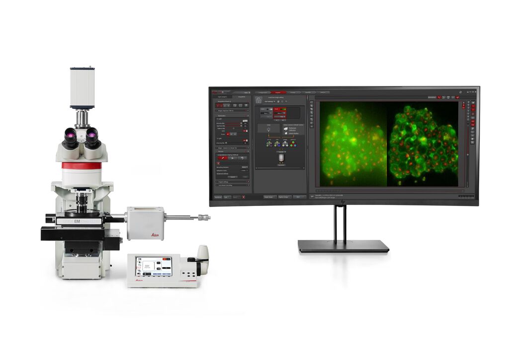
THUNDER Imager EM Cryo CLEM

Compreensão aprofundada da biologia estrutural celular

Leia os nossos artigos mais recentes

How to Successfully Perform Live-cell CLEM

The Leica Nano workflow provides a streamlined live-cell CLEM solution for getting insight bout structural changes of cellular components over time. Besides the technical handling described in the…

How to Successfully Implement Coral Life

The live-cell  CLEM workflow allows you to capture dynamic information related to a relevant biological process as it happens and put these observations into their ultrastructural context. The Leica…

Advancing Cellular Ultrastructure Research

Freeze-fracture and freeze-etching are useful tools for studying flexible membrane-associated structures such as tight junctions or the enteric glycocalyx. Freeze-fracture and etching are two…

The Cryo-CLEM Journey

This article describes the Cryo-CLEM technology and the benefits it can provide for scientists. Additionally, some scientific publications are highlighted. Recent developments in cryo electron…
3D reconstruction of an intercellular bridge in a C. elegans embryo

Download EM Workflow Solutions Booklet

This publication is a compilation of appropriate workflows for the most frequently used sample preparation methods, like Correlative Methodologies, Optogenetics & Electro-Physiology, Surface Analysis,…

Exploring the Structure and Life Cycle of Viruses

The SARS-CoV-2 outbreak started in late December 2019 and has since reached a global pandemic, leading to a worldwide battle against COVID-19. The ever-evolving electron microscopy methods offer a…
Cryo FIB lamella - Overlay of SEM and confocal fluorescence image. Target structure in yeast cells (nuclear pore proteine Nup159-Atg8-split Venus, red) marked by an arrow. Scale bar: 5 µm. Alegretti et al.,  Nature 586, 796-800 (2020).

Targeting Active Recycling Nuclear Pore Complexes using Cryo Confocal Microscopy

In this article, how cryo light microscopy and, in particular cryo confocal microscopy, is used to improve the reliability of cryo EM workflows is described. The quality of the EM grids and samples is…

Advancing Cell Biology with Cryo-Correlative Microscopy

Correlative light and electron microscopy (CLEM) advances biological discoveries by merging different microscopes and imaging modalities to study systems in 4D. Combining fluorescence microscopy with…

Workflows and Instrumentation for Cryo-electron Microscopy

Cryo-electron microscopy is an increasingly popular modality to study the structures of macromolecular complexes and has enabled numerous new insights in cell biology. In recent years, cryo-electron…
Mouse retina was fixed and stained by following reagents: anti-CD31 antibody (green): Endothelia cells, IsoB4 (red): Blood vessels, and microglia anti-GFAP antibody (blue): Astrocytes Sample courtesy by Jeremy Burton, PhD and Jiyeon Lee, PhD, Genentech Inc., South San Francisco, USA. Imaged by Olga Davydenko, PhD (Leica). Imaged with a THUNDER Imager 3D Cell Culture.

An Introduction to Computational Clearing

Many software packages include background subtraction algorithms to enhance the contrast of features in the image by reducing background noise. The most common methods used to remove background noise…

Crystal Clear Cryo Light-microscopy Images

This article describes how computational clearing of cryo light microscopy images improves the identification of cellular targets for cryo electron-microscopy.

Real Time Images of 3D Specimens with Sharp Contrast Free of Haze

THUNDER Imagers deliver in real time images of 3D specimens with sharp contrast, free of the haze or out-of-focus blur typical of widefield systems. They can even image clearly places deep inside a…

Campos de aplicação

Microscopia de fluorescência

A fluorescência é um dos fenômenos físicos mais comumente usados na microscopia biológica e analítica, principalmente devido a sua alta sensibilidade e alta especificidade. A fluorescência é uma forma…

Pesquisa de câncer

O câncer é uma doença complexa e heterogênea causada por células com deficiência na regulação do crescimento. Mudanças genéticas e epigenéticas em uma ou em um grupo de células prejudicam o…

Biologia celular

Quando a sua pesquisa é focada na compreensão da base celular da saúde e das doenças humanas, é crucial investigar as células de interesse em detalhes espaço-temporais e moleculares. Consequentemente,…

Virology

Soluções para aquisição de imagens e preparação de amostras para pesquisas em virologia

Organismos modelo na pesquisa

Um organismo modelo é uma espécie usada pelos pesquisadores para estudar processos biológicos específicos. Eles têm características genéticas similares aos seres humanos e são usados tipicamente em…

Tomografia crioeletrônica

A tomografia crioeletrônica (CryoET) é usada para resolver biomoléculas dentro de seu ambiente celular até uma resolução sem precedentes, abaixo de um nanômetro.

Técnicas avançadas de microscopia

As técnicas avançadas de microscopia abrangem tanto técnicas de imagem de alta resolução quanto de super-resolução. Estas técnicas são usadas principalmente para visualizar eventos biológicos em…

Microscopia correlativa de luz e eletrônica (CLEM)

Os fluxos de trabalho Coral da Leica Microsystems ajudam os usuários a correlacionar dados de microscopia de fluorescência e eletrônica (CLEM).
Background image
Scroll to top