STELLARIS Cryo
공초점 레이저 현미경
제품소개
홈
Leica Microsystems
STELLARIS Cryo 공초점 광학 현미경
최신 기사를 읽어 보세요

New Imaging Tools for Cryo-Light Microscopy
New cryo-light microscopy techniques like LIGHTNING and TauSense fluorescence lifetime-based tools reveal structures for cryo-electron microscopy.

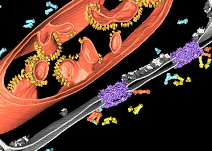
How to Target Fluorescent Structures in 3D for Cryo-FIB Milling
This article describes the major steps of the cryo-electron tomography workflow including super-resolution cryo-confocal microscopy. We describe how subcellular structures can be precisely located in…

Precise 3D Targeting for EM Imaging - Access What Matters
Find out how the seamless cryo-electron tomography workflow Coral Cryo uses confocal super resolution to target your structure of interest more precisely.

The Cryo-CLEM Journey
This article describes the Cryo-CLEM technology and the benefits it can provide for scientists. Additionally, some scientific publications are highlighted.
Recent developments in cryo electron…

Targeting Active Recycling Nuclear Pore Complexes using Cryo Confocal Microscopy
In this article, how cryo light microscopy and, in particular cryo confocal microscopy, is used to improve the reliability of cryo EM workflows is described. The quality of the EM grids and samples is…

Advancing Cell Biology with Cryo-Correlative Microscopy
Correlative light and electron microscopy (CLEM) advances biological discoveries by merging different microscopes and imaging modalities to study systems in 4D. Combining fluorescence microscopy with…

Crystal Clear Cryo Light-microscopy Images
This article describes how computational clearing of cryo light microscopy images improves the identification of cellular targets for cryo electron-microscopy.
Improve Cryo Electron Tomography Workflow
Leica Microsystems and Thermo Fisher Scientific have collaborated to create a fully integrated cryo-tomography workflow that responds to these research needs: Reveal cellular mechanisms at…
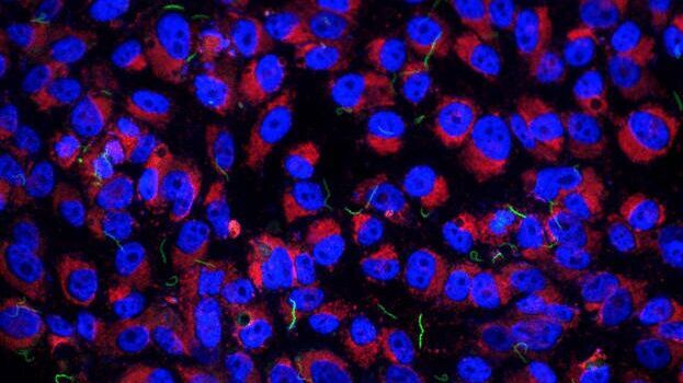
Imaging of Host Cell-bacteria Interactions using Correlative Microscopy under Cryo-conditions
Pathogenic bacteria have developed intriguing strategies to establish and promote infections in their respective hosts. Most bacterial pathogens initiate infectious diseases by adhering to host cells…
적용 분야
극저온 전자 단층 촬영술
극저온 전자 단층 촬영술(CryoET)는 세포 환경 내의 생체 분자를 1 nm 이하의 엄청난 해상도로 관찰하는 데 사용됩니다.
고급 현미경 기술
고급 현미경 기술에는 고해상도 및 초고해상도 이미징 기술이 모두 포함됩니다. 이러한 기술은 일반적으로 세포 또는 조직인 시료에 대해 가능한 한 부드럽게 생물학적 이벤트를 매우 높은 해상도로 시각화하는 데 주로 사용됩니다. 연구자들은 첨단 현미경 기술의 도움을 받아 생물학적 경로, 유전자 또는 단백질 발현, 질병 메커니즘 등에 중대한 영향을 미치는 생체…
상관관계적 광학 현미경과 전자현미경(CLEM)
라이카마이크로시스템즈 Coral 작업 흐름은 사용자가 형광 현미경과 전자현미경 (CLEM) 데이터의 상관관계를 분석하는 데 도움이 됩니다.
