

Ciências da vida
Ciências da vida
Este é o lugar para expandir seus conhecimentos, recursos de pesquisa e aplicações práticas de microscopia em vários campos científicos. Saiba como obter visualização precisa, interpretação de imagens e avanços na pesquisa. Encontre informações perspicazes sobre microscopia avançada, técnicas de geração de imagens, preparação de amostras e análise de imagens. Os tópicos abordados incluem biologia celular, neurociência e pesquisa do câncer, com foco em aplicações e inovações de ponta.
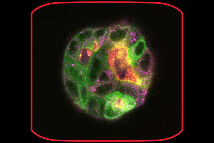
How To Perform Fast & Stable Multicolor Live-Cell Imaging
With the help of live-cell imaging researchers gain insights into dynamic processes of living cells up to whole organisms. This includes intracellular as well as intercellular activities. Protein or…
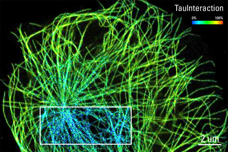
TauInteraction – Studying Molecular Interactions with TauSense
Fluorescence microscopy constitutes one of the pillars in life sciences and is a tool commonly used to unveil cellular structure and function. A key advantage of fluorescence microscopy resides in the…
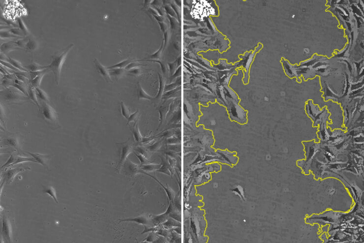
Studying Wound Healing of Smooth Muscle Cells
This article discusses how wound healing of cultured smooth muscle cells (SMCs) in multiwell plates can be reliably studied over time with less effort using a specially configured Leica inverted…
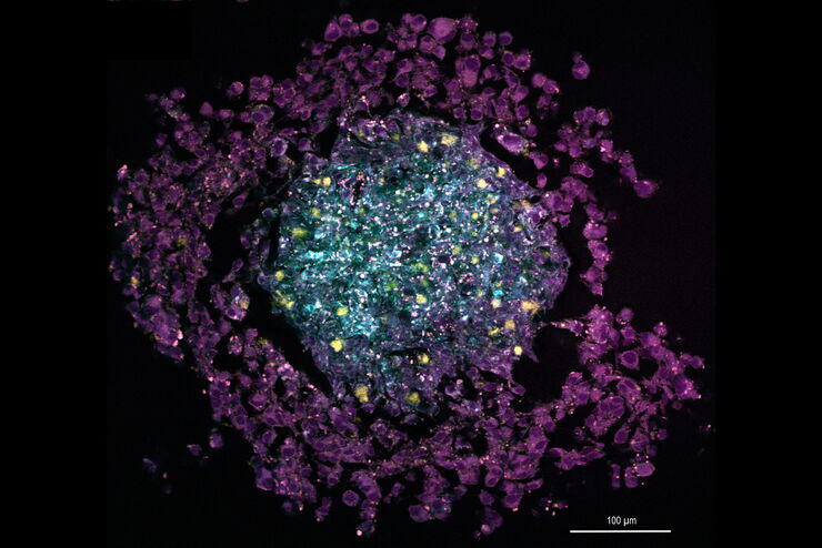
The Potential of Coherent Raman Scattering Microscopy at a Glance
Coherent Raman scattering microscopy (CRS) is a powerful approach for label-free, chemically specific imaging. It is based on the characteristic intrinsic vibrational contrast of molecules in the…
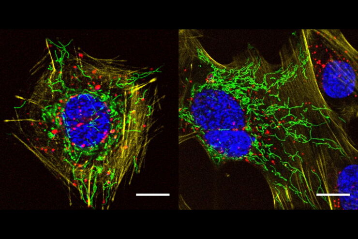
A Versatile Palette of Fluorescent Probes
Researchers at the Max Planck Institute for Medical Research in Heidelberg have developed a general strategy to synthesize live-cell compatible fluorogenic probes, and the result are the new MaP (Max…
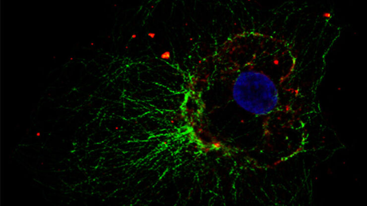
How to Prepare your Specimen for Immunofluorescence Microscopy
Immunofluorescence (IF) specimen preparations can be analyzed by various fluorescence microscopy techniques, depending on the research application. IF is indispensable for researchers using a simple…
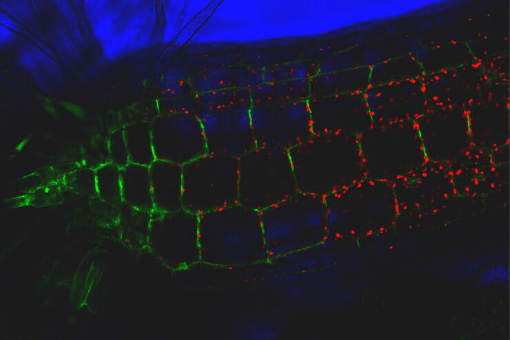
Live-Cell Imaging Techniques
The understanding of complex and/or fast cellular dynamics is an important step for exploring biological processes. Therefore, today’s life science research is increasingly focused on dynamic…

Imaging of Anti-Cancer Drug Uptake in Spheroids using DLS
Spheroid 3D cell culture models mimic the physiology and functions of living tissues making them a useful tool to study tumor morphology and screen anti-cancer drugs. The drug AZD2014 is a recognized…

Artificial Intelligence and Confocal Microscopy – What You Need to Know
This list of frequently asked questions provides “hands-on” answers and is a supplement to the introductory article about Dynamic Signal Enhancement powered by Aivia "How Artificial Intelligence…


