Contacto
Contacto

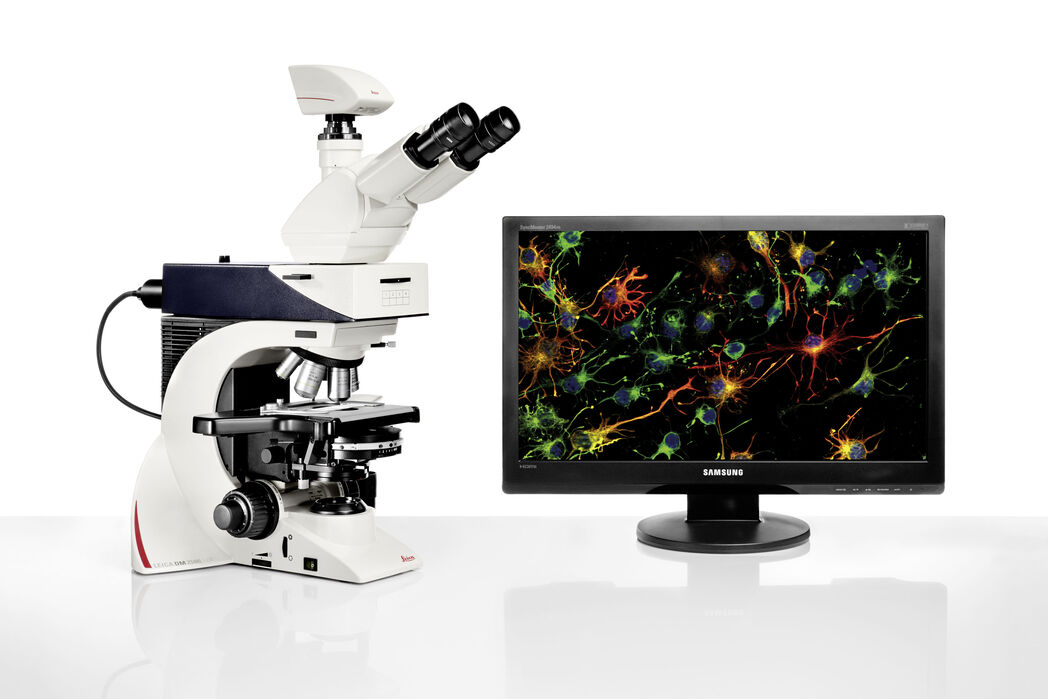
Leica DM2500 & DM2500 LED Documentación sencilla y económica

Capture y procese hoy mismo imágenes con fluorescencia y grandes prestaciones adaptadas a sus necesidades y presupuesto

Lea nuestros últimos artículos

Staining is crucial to revealing details in the specimens.

H&E Staining in Microscopy

If we consider the role of microscopy in pathologists’ daily routines, we often think of the diagnosis. While microscopes indeed play a crucial role at this stage of the pathology lab workflow, they…
Acute lymphoblastic leukemia

How to Benefit from Digital Cytopathology

If you have thought of digital cytopathology as characterized by the digitization of glass slides, this webinar with Dr. Alessandro Caputo from the University Hospital of Salerno, Italy will broaden…
Laboratory

Factors for Selecting Clinical Microscopes

If you are considering the purchase of new clinical microscopes for yourself or your team, you will think about many options, vendors, and microscope models. Here are some tips to help you come to the…
H&E stained specimen, 20x magnification

The Time to Diagnosis is Crucial in Clinical Pathology

Abnormalities in tissues and fluids - that’s what pathologists are looking for when they examine specimens under the microscope. What they see and deduce from their findings is highly influential, as…
Steps of Koehler Illumination

Koehler Illumination: A Brief History and a Practical Set Up in Five Easy Steps

In this article, we will look at the history of the technique of Koehler Illumination in addition to how to adjust the components in five easy steps.
SPERM HY-LITER™ staining of a sexual assault smear slide performed by forensic DNA crime laboratory.

Forensic Detection of Sperm from Sexual Assault Evidence

The impact of modern scientific methods on the analysis of crime scene evidence has dramatically changed many forensic sub-specialties. Arguably one of the most dramatic examples is the impact of…

Campos de aplicación

Microscopía de fluorescencia

La fluorescencia es uno de los fenómenos físicos más utilizados en la microscopía biológica y analítica, principalmente por su alta sensibilidad y alta especificidad. Averigüe cómo los microscopios de…

Patología clínica

Descubra cómo los microscopios ergonómicos de Leica Microsystems permiten realizar diagnósticos precisos y oportunos en patología clínica.

Microscopio para patología

En ocasiones, el análisis de especímenes patológicos requiere muchas horas de trabajo frente a un microscopio. Como consecuencia, el usuario puede sentir molestias físicas y tensión que puede conducir…

Patología anatómica

Descubra cómo los microscopios ergonómicos de Leica Microsystems facilitan un diagnóstico eficiente y preciso en la patología anatómica.

Técnicas básicas de microscopía

Se utilizan técnicas básicas de microscopía en las que todo el espécimen en la platina del microscopio se expone a una fuente de luz. Todo el espécimen se ilumina con luz blanca desde arriba (en una…

Microscopios DIC

Un microscopio DIC es un microscopio de campo ancho que tiene un filtro de polarización y un prisma Wollaston entre la fuente de luz y la lente del condensador, así como entre la lente del objetivo y…

Microscopios de campo oscuro

El método de contraste de campo oscuro aprovecha la difracción o dispersión de la luz de las estructuras de un espécimen biológico o las características no uniformes de una muestra de material.
Background image
Scroll to top