Productos

Productos

Productos

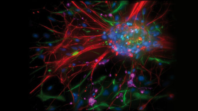
Desarrollamos nuestros sistemas de precisión de tecnología punta para el análisis de microestructuras con el usuario, para el usuario. En nuestra cartera de productos encontrará soluciones de sistema para ciencias biológicas, incluyendo la biotecnología y la medicina, así como la investigación y el desarrollo de materias primas y la garantía de la calidad industrial.

Productos

Nuestras Últimas Novedades

Echa un vistazo a todas nuestras próximas conferencias, congresos, ferias de muestras, webinarios y talleres. Únete a nosotros en uno de nuestros próximos eventos!

Todos los eventos

Nuestros últimos artículos

El portal de conocimiento de Leica Microsystems ofrece investigación científica y material didáctico sobre temas de microscopía. El contenido está diseñado para apoyar a principiantes, profesionales experimentados y científicos por igual en su trabajo diario y experimentos.

Más artículos

Investigación con Pez Cebra

Para obtener los mejores resultados en clasificación, selección, manipulación y captura y procesamiento de imágenes necesita ver los detalles y estructuras para poder tomar las decisiones adecuadas con respecto a los siguientes pasos de la investigación. Famosos por una óptica sobresaliente y una excelente resolución, los microscopios estereoscópicos y las bases de diascopía Leica se han convertido en la opción favorita de investigadores de todo el mundo.

The Guide to Augmented Reality in Microsurgery

In an era of technological advancement, Augmented Reality (AR) is rapidly transforming the medical field. In surgical microscopy, AR can display fluorescence signals as digital overlays in real-time within the surgical field of view, providing surgeons with additional information about fluorescent areas of interest. A surgeon's view can be augmented with data overlays from different systems, offering functional or anatomical information superimposed onto the live microscope view.

How Fluorescence Guides Sectioning of Resin-embedded EM Samples

Electron microscopes, including transmission electron microscopes (TEM) and scanning electron microscopes (SEM), are widely utilized to gain detailed structural information about biological samples or non-living materials. Ultramicrotomy is the preferred technique for producing ultrathin sections, less than 100 nm thick for TEM/SEM analysis. During sample preparation small sample pieces are embedded in epoxy or acrylic resin, excess resin is trimmed away, and the specimen is sliced into ultrathin sections (50 nm - 100 nm) using a glass or diamond knife.

Coherent Raman Scattering Microscopy Publication List

CRS (Coherent Raman Scattering) microscopy is an umbrella term for label-free methods that image biological structures by exploiting the characteristic, intrinsic vibrational contrast of their molecules. The two most important CRS techniques are Coherent Anti-Stokes Raman Scattering (CARS) and Stimulated Raman Scattering (SRS). The biochemical image contrast of CRS is in many ways complementary to the molecular contrast obtained in fluorescence microscopy. A second crucial advantage of these methods is that they preserve the specimen/sample in a near pristine state. This reference list presents current and basic papers on CRS microscopy.

Microscopios para disección

Las disecciones pueden implicar pasar horas mirando por los oculares de un microscopio para disección. Leica Microsystems le permite escoger entre una variada gama de microscopios y un amplio rango de piezas y accesorios para microscopios de disección, para que pueda encontrar la solución que mejor se adapte a sus necesidades.

A Guide to Polarized Light Microscopy

Polarized light microscopy (POL) enhances contrast in birefringent materials and is used in geology, biology, and materials science to study minerals, crystals, fibers, and plant cell walls.

Factors to Consider when Selecting Clinical Microscopes

What matters if you would like to purchase a clinical microscope? Learn how to arrive at the best buying decision from our Science Lab Article.

Clinical Microscopy: Considerations on Camera Selection

The need for images in pathology laboratories has significantly increased over the past few years, be it in histopathology, cytology, hematology, clinical microbiology, or other applications. They serve many purposes on top of the documentation of the diagnosis. Yet, the view through the eyepieces and the image are different in nature, the one is an optical image, the other a digital image. Looking at a few aspects of this process that are related cameras will help you make sure you can obtain the images with all the detail and color fidelity you need.
Scroll to top