Especialidades médicas
Especialidades médicas
Explore uma coleção abrangente de recursos científicos e clínicos adaptados para profissionais de saúde, incluindo percepções de colegas, estudos de casos clínicos e simpósios. Projetado para neurocirurgiões, oftalmologistas e especialistas em cirurgia plástica e reconstrutiva, otorrinolaringologia e odontologia. Esta coleção destaca os mais recentes avanços em microscopia cirúrgica. Descubra como as tecnologias cirúrgicas de ponta, como fluorescência AR, visualização 3D e imagens intraoperatórias de OCT, possibilitam a tomada de decisões confiantes e a precisão em cirurgias complexas.
Guide to Live-Cell Imaging
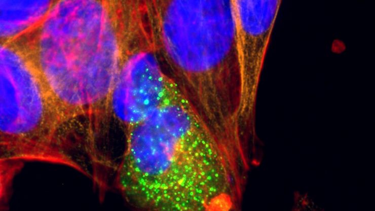
For a wide range of applications in various research fields of life science, live-cell imaging is an indispensable tool for visualizing cells in a state as close to in vivo, i.e. living and active, as…
Microscopy and AI Solutions for 2D Cell Culture
This eBook explores the integration of microscopy and AI technologies in 2D cell culture workflows. It highlights how traditional imaging methods—such as brightfield, phase contrast, and…
Faster & Deeper Insights into Organoid and Spheroid Models
Gain deeper, more translatable, insights into organoid and spheroid models for drug discovery and disease research by overcoming key imaging challenges. In this eBook, explore advanced microscopy…
A Microvascular Surgeon’s View: How MyVeo Transforms Visualization
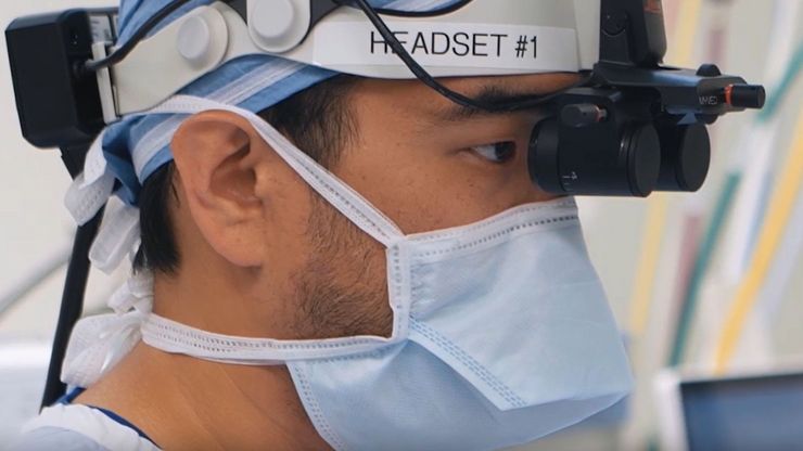
In this article, Dr. Andrew T. Huang, MD, FACS, otolaryngologist and a head and neck reconstructive surgeon, shares how digital 3D surgical visualization with the MyVeo headset from Leica Microsystems…
How to Image Axon Regeneration in Deep Muscle Tissue
This study highlights Dr. Aaron Lee’s research on mapping nerve regeneration in muscle grafts post-amputation. Limb loss often leads to reduced quality of life, not only from tissue loss but also due…
How to Select the Right Measurement Microscope
With a measurement microscope, users can measure the size and dimensions of sample features in both 2D and 3D, something crucial for inspection, QC, failure analysis, and R&D. However, choosing the…
Microscope Calibration for Measurements: Why and How You Should Do It
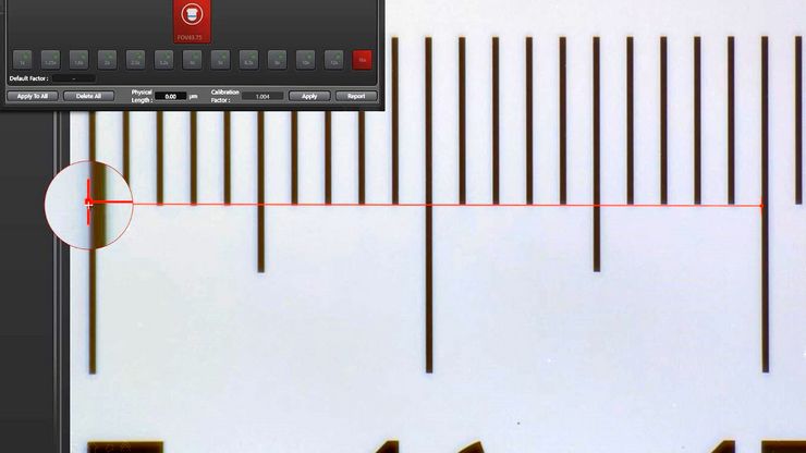
Microscope calibration ensures accurate and consistent measurements for inspection, quality control (QC), failure analysis, and research and development (R&D). Calibration steps are described in this…
Neurocientífica
Você está trabalhando rumo a uma melhor compreensão de doenças neurodegenerativas ou estudando a função do sistema nervoso? Veja como você pode fazer descobertas com as soluções de aquisição de…
The Guide to Augmented Reality in Microsurgery
In an era of technological advancement, Augmented Reality (AR) is rapidly transforming the medical field. In surgical microscopy, AR can display fluorescence signals as digital overlays in real-time…


