STELLARIS 5 Cryo
Microscopios confocales
Productos
Inicio
Leica Microsystems
STELLARIS 5 Cryo Microscopio óptico confocal
Lea nuestros últimos artículos
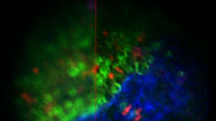
New Imaging Tools for Cryo-Light Microscopy
New cryo-light microscopy techniques like LIGHTNING and TauSense fluorescence lifetime-based tools reveal structures for cryo-electron microscopy.
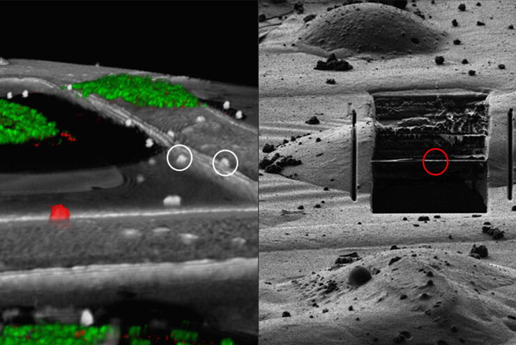
How to Target Fluorescent Structures in 3D for Cryo-FIB Milling
This article describes the major steps of the cryo-electron tomography workflow including super-resolution cryo-confocal microscopy. We describe how subcellular structures can be precisely located in…
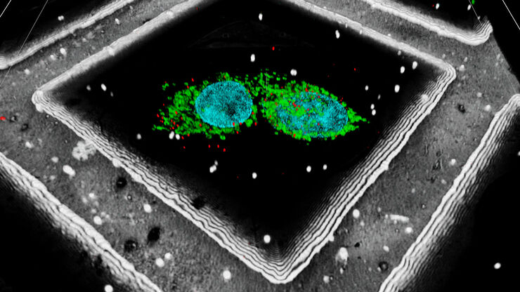
Precise 3D Targeting for EM Imaging - Access What Matters
Find out how the seamless cryo-electron tomography workflow Coral Cryo uses confocal super resolution to target your structure of interest more precisely.
Campos de aplicación
Tomografía crioelectrónica
La tomografía crioelectrónica (CryoET) se utiliza para resolver biomoléculas dentro de su entorno celular hasta una resolución sin precedentes por debajo de un nanómetro.
Técnicas avanzadas de microscopía
Las técnicas de microscopía avanzada abarcan tanto las técnicas de imagen de alta resolución como las de superresolución. Estas técnicas se utilizan principalmente para visualizar eventos biológicos…
Microscopía correlativa óptica y electrónica (CLEM)
Los flujos de trabajo Coral de Leica Microsystems ayudan a los usuarios a correlacionar imágenes de microscopía de fluorescencia y microscopía electrónica (CLEM).


