Contacto
Contacto

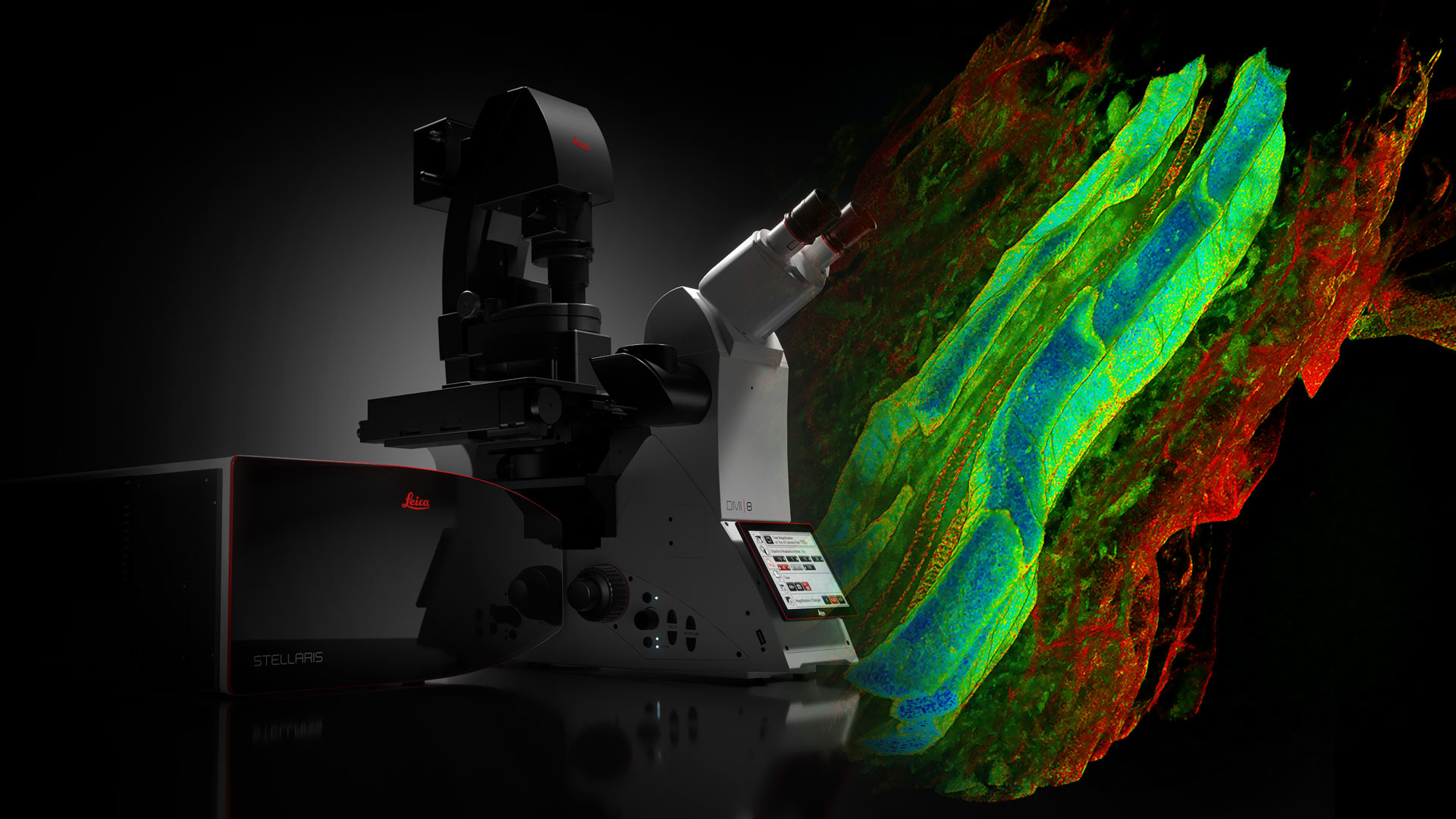
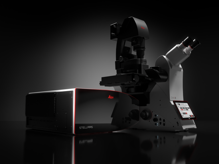
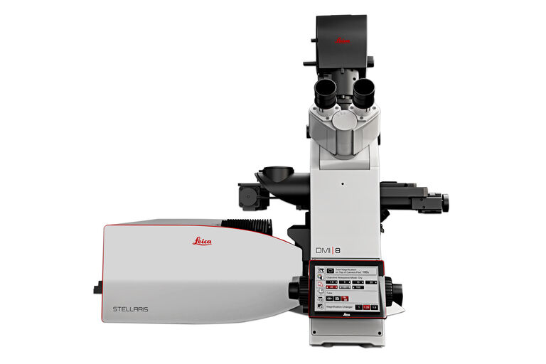
STELLARIS 5 & STELLARIS 8 Sistemas de microscopía confocal

Power. Potential. Productivity.

Lea nuestros últimos artículos

Murine esophageal organoids (DAPI, Integrin26-AF 488, SOX2-AF568) imaged with the THUNDER Imager 3D Cell Culture. Courtesy of Dr. F.T. Arroso Martins, Tamere University, Finland.

How to Get Deeper Insights into your Organoid and Spheroid Models

In this eBook, learn about key considerations for imaging 3D cultures, such as organoids and spheroids, and discover microscopy solutions to shed new insights into dynamic processes in 3D real-time
Fluorescence microscopy image of liver tissue where DNA in the nuclei are stained with Feulgen-pararosanilin and visualized with transmitted green light.

Epi-Illumination Fluorescence and Reflection-Contrast Microscopy

This article discusses the development of epi-illumination and reflection contrast for fluorescence microscopy concerning life-science applications. Much was done by the Ploem research group…
Application example of hyperspectral imaging

Potential of Multiplex Confocal Imaging for Cancer Research and Immunology

Explore the new frontiers of multi-color fluorescent imaging: from image acquisition to analysis
Molecular structure of the green fluorescent protein (GFP)

Introduction to Fluorescent Proteins

Overview of fluorescent proteins (FPs) from, red (RFP) to green (GFP) and blue (BFP), with a table showing their relevant spectral characteristics.
Multi-tissue array with 4 markers shown including DAPI, NaKATPase, PanCk, and Vimentin.

Spatial Biology: Learning the Landscape

Spatial Biology: Understanding the organization and interaction of molecules, cells, and tissues in their native spatial context
In vivo imaging of a mouse pial and cortical vasculature through a glass window (ROSAmT/mG::Pdgfb-CreERT2 mouse meningeal and cortical visualization following tamoxifen induction and craniotomy). Courtesy: Thomas Mathivet, PhD

Windows on Neurovascular Pathologies

Discover how innate immunity can sustain deleterious effects following neurovascular pathologies and the technological developments enabling longitudinal studies into these events.
Lifetime-based multiplexing in live cells using TauSeparation. Mammalian cells expressing LifeAct-GFP (ibidi GmbH) and labelled with MitoTracker Green. Acquisition with one detector, intensity information shown in grey. The two markers can be separated using lifetime information: LifeAct-GFP (cyan), MitoTracker Green (magenta). Image acquired with STELLARIS 5.

The Power of Reproducibility, Collaboration and New Imaging Technologies

In this webinar you willl learn what impacts reproducibility in microscopy, what resources and initiatives there are to improve education and rigor and reproducibility in microscopy and how…
AI-based workflow for fast rare event detection in living biological samples using Autonomous Microscopy powered by Aivia

AI Microscopy Enables the Efficient Detection of Rare Events

Localization and selective imaging of rare events is key for the investigation of many processes in biological samples. Yet, due to time constraints and complexity, some experiments are not feasible…
How is microscopy used in spatial biology - Teaserimage

How is Microscopy Used in Spatial Biology? A Microscopy Guide

Different spatial biology methods in microscopy, such as multiplex imaging, are helping to better understand tissue landscapes. Learn more in this microscopy guide.
Five-color FLIM-STED

Five-color FLIM-STED with One Depletion Laser

Webinar on five-color STED with a single depletion laser and fluorescence lifetime phasor separation.

Confocal Imaging of Immune Cells in Tissue Samples

In this webinar, you will discover how to perform 10-color acquisition using a confocal microscope. The challenges of imaged-based approaches to identify skin immune cells. A new pipeline to assess…

Virtual Reality Showcase for STELLARIS Confocal Microscopy Platform

In this webinar, you will discover how to perform 10-color acquisition using a confocal microscope. The challenges of imaged-based approaches to identify skin immune cells. A new pipeline to assess…
Combining spectrally resolved detection and fluorescence lifetime multiplexing

Live-Cell Fluorescence Lifetime Multiplexing Using Organic Fluorophores

On-demand video: Imaging more subcellular targets by using fluorescence lifetime multiplexing combined with spectrally resolved detection.
Donor (D) and acceptor (A) molecule which participate in FRET (Förster resonance energy transfer).

What is FRET with FLIM (FLIM-FRET)?

This article explains the FLIM-FRET method which combines resonance energy transfer and fluorescence lifetime imaging to study protein-protein interactions.

Insights into Vesicle Trafficking

STELLARIS provides integral access to complementary layers of information for dynamic, structural, and mechanistic insights into vesicle trafficking.

Visualizing Protein-Protein Interactions by Non-Fitting and Easy FRET-FLIM Approaches

The Webinar with Dr. Sergi Padilla-Parra is about visualizing protein-protein interaction. He gives insight into non-fitting and easy FRET-FLIM approaches.
Spectral separation of 11 fluorophores coupled to polystyrene beads on a STELLARIS confocal system.

Multiplexing through Spectral Separation of 11 Colors

Fluorescence microscopy is a fundamental tool for life science research that has evolved and matured together with the development of multicolor labeling strategies in cells tissues and model…
Transverse histological cut of a rabbit tongue. 50 Mpixels images (2326 µm x 1739 µm) in 14 x 18 tiles. Lifetime gives an additional contrast that allows to differentiate different structures in histological stainings.

A Guide to Fluorescence Lifetime Imaging Microscopy (FLIM)

The fluorescence lifetime is a measure of how long a fluorophore remains on average in its excited state before returning to the ground state by emitting a fluorescence photon.

TauInteraction – Studying Molecular Interactions with TauSense

Fluorescence microscopy constitutes one of the pillars in life sciences and is a tool commonly used to unveil cellular structure and function. A key advantage of fluorescence microscopy resides in the…
Identification of distinct structures_roundworm_Ascaris_female

Find Relevant Specimen Details from Overviews

Switch from searching image by image to seeing the full overview of samples quickly and identifying the important specimen details instantly with confocal microscopy. Use that knowledge to set up…
Dynamic Signal Enhancement powered by Aivia:  Truly simultaneous multicolor imaging of live cells (U2OS) in 3D

Artificial Intelligence and Confocal Microscopy – What You Need to Know

This list of frequently asked questions provides “hands-on” answers and is a supplement to the introductory article about Dynamic Signal Enhancement powered by Aivia "How Artificial Intelligence…
Dynamic Signal Enhancement powered by Aivia: Truly simultaneous multicolor imaging of live cells (U2OS) in 3D

How Artificial Intelligence Enhances Confocal Imaging

In this article, we show how artificial intelligence (AI) can enhance your imaging experiments. Namely, how Dynamic Signal Enhancement powered by Aivia improves image quality while capturing the…

Spectroscopic Evaluation of Red Blood Cells

Hemoglobinopathies are a major healthcare problem. This study presents a possible diagnostic tool for thalassemia which is based on confocal spectroscopy. This approach exploits spectral detection and…

Visualizing Protein Degradation and Aggregation in the Living Cell

Our guest speaker, Prof Dr Eric Reits, presents his work on neurodegenerative disorders. Reits’ group are experts on the subject of Huntington’s disease and work towards identifying leads for…

Life Beyond the Pixels: Deep Learning Methods for Single Cell Analysis

Our guest speaker Prof Dr Peter Horvath presents his work on single cell-based large-scale microscopy experiments. This novel targeting approach includes the use of machine learning models and…
Nematostella

Live Cell Imaging Gallery

Live cell microscopy techniques are fundamental to get a better understanding of cellular and molecular function. Today, widefield microscopy is the most common technique used to visualize cell…

Super-Resolution Microscopy Image Gallery

Due to the diffraction limit of light, traditional confocal microscopy cannot resolve structures below ~240 nm. Super-resolution microscopy techniques, such as STED, PALM or STORM or some…

Tissue Image Gallery

Visual analysis of animal and human tissues is critical to understand complex diseases such as cancer or neurodegeneration. From basic immunohistochemistry to intravital imaging, confocal microscopy…
Virally labeled neurons (red) and astrocytes (green) in a cortical spheroid derived from human induced pluripotent stem cells. THUNDER Model Organism Imagerwith a 2x 0.15 NA objective at 3.4x zoomwas used to produce this 425 μm Z-stack (26 positions), which is presented here as an Extended Depth of Field(EDoF)projection.

Neuroscience Images

Neuroscience commonly uses microscopy to study the nervous system’s function and understand neurodegenerative diseases.

Multicolor Image Gallery

Fluorescence multicolor microscopy, which is one aspect of multiplex imaging, allows for the observation and analysis of multiple elements within the same sample – each tagged with a different…

Cancer Research Image Gallery

Fluorescence microscopy allows the study of changes occurring in tissue and cells during cancer development and progression. Techniques such as live cell imaging are critical to understand cancer…

Cell Biology Image Gallery

Cell biology studies the structure, function and behavior of cells, including cell metabolism, cell cycle, and cell signaling. Fluorescence microscopes are an integral part of a cell biologist…

Adding Dimensions to Multiplex Molecular Imaging

Molecular imaging of living specimens offers a means to draw upon the growing body of high-throughput molecular data to better understand the underlying cellular and molecular mechanisms of complex…
Root-hypocotyl junction of Arabidopsis thaliana. Image acquired with TauContrast. Sample courtesy: Dr. Melanie Krebs, COS, University of Heidelberg.

Benefits of TauContrast to Image Complex Samples

In this interview, Dr. Timo Zimmermann talks about his experience with the application of TauSense tools and their potential for the investigation of demanding samples such as thick samples or…
Influenca in lung epithelial cells (porcine) - THUNDER Imager 3D Cell Culture Influenca virus – red, cilia – green, Nuclei – blue.

How Can Immunofluorescence Aid Virology Research?

Modern virology research has become as crucial now as ever before due to the global COVID-19 pandemic. There are many powerful technologies and assays that virologists can apply to their research into…

Obtenga el máximo de información de su muestra con LIGHTNING

LIGHTNING es un proceso adaptativo para la extracción de información que revela estructuras finas y detalles que de otro modo simplemente no serían visibles, de forma completamente automática . A…

Explore Innovative Techniques to Separate Fluorophores with Overlapping Spectra

In this article we explore several strategies you can take to improve the separation of fluorophores and increase the number of fluorescent probes you can distinguish in your sample.

STELLARIS White Light Lasers

When it comes to choosing fluorescent probes for your multi-color experiments, you shouldn’t have to compromise. Now you can advance beyond conventional excitation sources that limit your fluorophore…

TauSense Technology Imaging Tools

Leica Microsystems’ TauSense technology is a set of imaging modes based on fluorescence lifetime. Found at the core of the STELLARIS confocal platform, it will revolutionize your imaging experiments.…

The Power HyD Detector Family

Powerful photon counting detectors on the STELLARIS confocal platform provide improved photon counting, ultra-sensitive imaging and more color options in the NIR spectrum.
Fluorescence microscopy image on the left with no distinction between the fluorescent signal and background autofluorescence. FLIM was used in the image on the right to differentiate autofluorescence in chloroplasts (blue) from the desired fluorescent signal from the cell membrane (green).

Learn how to Remove Autofluorescence from your Confocal Images

Autofluorescence can significantly reduce what you can see in a confocal experiment. This article explores causes of autofluorescence as well as different ways to remove it, from simple media fixes to…

DIVE Multiphoton Microscope Image Gallery

Today’s life science research focusses on complex biological processes, such as the causes of cancer and other human diseases. A deep look into tissues and living specimens is vital to understanding…

Nobel Prize 2013 in Physiology or Medicine for Discoveries of the Machinery Regulating Vesicle Traffic

On October 7th 2013, The Nobel Assembly at Karolinska Institutet has decided to award The Nobel Prize in Physiology or Medicine 2012 jointly to James E. Rothman, Randy W. Schekman and Thomas C. Südhof…

Handbook of Optical Filters for Fluorescence Microscopy

Fluorescence microscopy and other light-based applications require optical filters that have demanding spectral and physical characteristics. Often, these characteristics are application-specific and…
Fluorescence microscope image of a life-science specimen

An Introduction to Fluorescence

This article gives an introduction to fluorescence and photoluminescence, which includes phosphorescence, explains the basic theory behind them, and how fluorescence is used for microscopy.

Campos de aplicación

Imagen de célula viva

Con un cambio de perspectiva desde componentes individuales del microscopio a una solución de captura y procesamiento de imágenes de células vivas totalmente operativa, Leica Microsystems integra…

Microscopios de súper-resolución y nanoscopios

Leica Microsystems es una empresa pionera en microscopía de superresolución, que trabaja en estrecha colaboración con científicos de primera línea en este campo. Leica Microsystems es el único…

Investigación del cáncer

El cáncer es una enfermedad compleja y heterogénea causada por células con una regulación deficiente del crecimiento. Los cambios genéticos y epigenéticos en una célula o en un grupo de ellas alteran…

Organoides y cultivos celulares en 3D

Uno de los avances recientes más emocionantes en la investigación de las ciencias biológicas es el desarrollo de sistemas de cultivo celular en 3D, como organoides, esferoides y modelos de órganos en…

Neurociencia

¿Trabaja para comprender mejor las enfermedades neurodegenerativas o la función del sistema nervioso? Vea cómo puede conseguir descubrimientos con las soluciones de imágenes de Leica Microsystems.

Microscopía de fluorescencia

La fluorescencia es uno de los fenómenos físicos más utilizados en la microscopía biológica y analítica, principalmente por su alta sensibilidad y alta especificidad. Averigüe cómo los microscopios de…

Fluorescence Lifetime Imaging

La microscopía de tiempo de vida de fluorescencia (FLIM) es una técnica que hace uso de las propiedades inherentes de las marcadores fluorescentes. Además de tener espectros de emisión…

Biología celular

Descubra cómo puede extraer el máximo rendimiento a sus investigaciones en materia de biología celular y amplíe sus descubrimientos con las soluciones de generación de imágenes de Leica Microsystems.

Virología

¿Su interés en investigación se centra en infecciones víricas y enfermedades? Descubra cómo puede adquirir conocimientos sobre virología con soluciones de generación de imágenes y preparación de…

Organismos modelo en la investigación

Un organismo modelo es una especia empleada por los investigadores para estudiar procesos biológicos específicos. Sus características genéticas son similares a las de los humanos y se emplean de forma…
Scroll to top