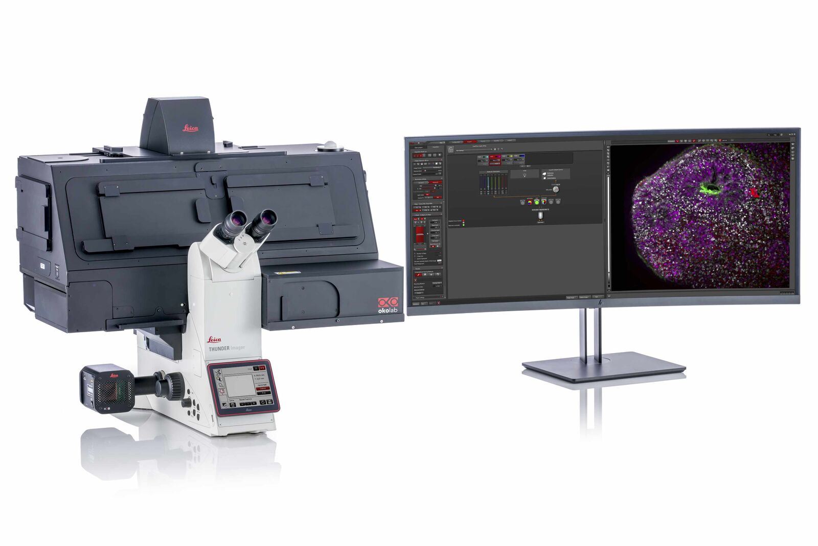
THUNDER Imager Live Cell & 3D Assay
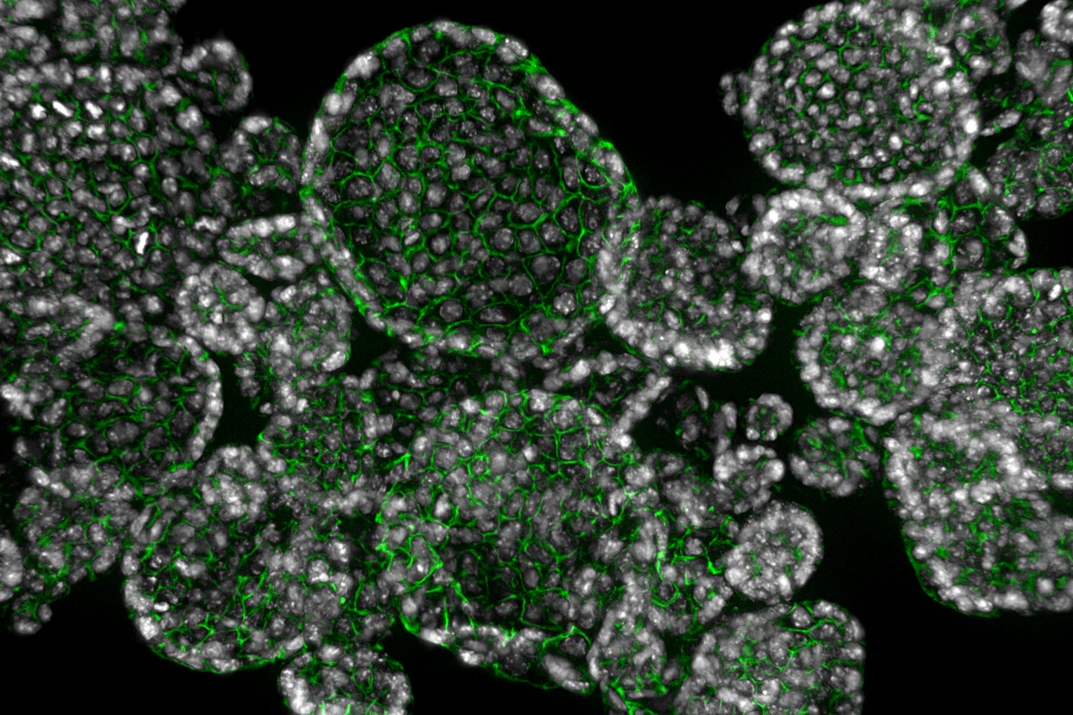
Les THUNDER Imagers vous offrent une solution pour l'analyse 3D de cultures de cellules complexes, que vous souhaitiez étudier les cellules souches, les sphéroïdes ou les organoïdes.
Les THUNDER Imagers intègrent « Computational Clearing », la technologie innovante de Leica. Elle élimine en toute efficacité le flou hors focus en temps réel, permettant ainsi d'utiliser judicieusement des échantillons 3D avec des microscopes à fluorescence avec une détection par caméra. La sensibilité élevée du système garantit une phototoxicité et un photoblanchiment faibles, en d'autres termes un plus haut débit avec des conditions optimales.
Haut débit pour de meilleures statistiques et un flux de travail efficace
Automatisez vos analyses 3D de cultures de cellules pour étudier efficacement les modèles de pathologies de demain. THUNDER vous permet d'imager des volumes d'échantillons importants, tels que des organoïdes pulmonaires, à grande vitesse. En outre, l'automatisation réduit au maximum l'interaction de l'utilisateur, même pour les expériences complexes.
Résultats :
- Données précises et fiables en moins de temps
- Plus haut débit
- Meilleures statistiques et meilleurs résultats
")

Conditions physiologiques optimales - exposition minimale à la lumière
En matière de culture cellulaire 3D, il est primordial d'observer la véritable physiologie pour obtenir des résultats significatifs. En règle générale, vous souhaitez étudier vos cellules pendant qu'elles se trouvent dans un état proche de l'état naturel en optimisant les conditions expérimentales, par exemple, l'intensité lumineuse la plus faible et les durées d'exposition les plus courtes possibles.
THUNDER Imager Live Cell répond à ces exigences avec une source LED haut de gamme, dotée d'une petite bande passante optimisée pour l'excitation. Même avec un faible éclairage et des durées d'exposition courtes, la caméra sCMOS sensible haut de gamme livre des informations d'image pertinentes grâce à une efficacité quantique de 82 % maximum.
Afin de réduire davantage l'exposition de l'échantillon à la lumière, l'éclairage est limité à la durée d'enregistrement réelle. L'obturateur de la caméra est synchronisé avec la source de lumière LED Lumencor à grande vitesse (durée de commutation de 20 µs) afin de réduire au maximum le photoblanchiment.
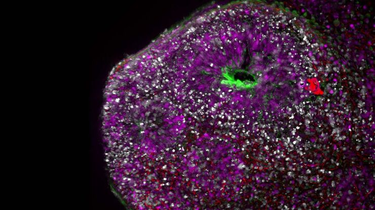
Développement du pancréas du poisson zèbre
Le système THUNDER Imager 3D Assay a permis d'identifier clairement les cellules alpha (GFP-vert) et bêta (mCardinal-rouge) dans un pancréas de poisson zèbre en développement.
Imagé dans les canaux bleu (Hoechst), vert (GFP) et rouge (mCardinal), cette image z-stack de 150 images a été complétée avec tous les canaux en une minute.
Les conditions physiologiques de l'échantillon sont maintenues en minimisant le photoblanchiment, en fournissant des images de haute qualité et un débit élevé de données, ce qui améliore l'efficacité du workflow.
Images fournies avec l'aimable autorisation de Radhan Ramadass et Yu Hsuan du Max Planck Institute for Heart and Lung Research, Bad Nauheim (Allemagne)


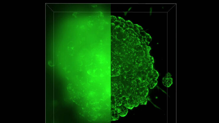
Image en temps réel – Processus cellulaires
Les mécanismes cellulaires sont très rapides, tout particulièrement pour une cellule. Les expériences d'imagerie de cellules vivantes les plus modernes exigent des systèmes à grande vitesse.
THUNDER Imager Live Cell vous apporte la puissance de systèmes à fluorescence par caméra sCMOS ultra-sensibles utilisant des images Full Frame en une seule prise.
Conjointement avec une sensibilité élevée, THUNDER Imager Live Cell vous permet de capturer des données jusqu'à 90 images par seconde, vous aidant ainsi à observer des événements cellulaires rapides. Ils capturent des données d'image nettes rapidement, même au cœur d'un amas cellulaire 3D épais. Vous pouvez demeurer au fait des processus rapides, même au cours d'expériences sur les longueurs d'onde d'émission multiples, grâce à des roues de filtres externes, très rapides d'une position à une autre (< 27 ms).
