Filter articles
Tags
Produits
Loading...

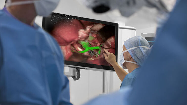
AR Fluorescence in Aneurysm Clipping and AVM Surgery
Discover how GLOW800 Augmented Reality fluorescence supports neurovascular surgical procedures and in particular aneurysm clipping and AVM surgery.
Loading...
Clinical Uses in Cerebrovascular and Skull Base Neurosurgery
In this webinar Dr. Bendok and Dr. Morcos explain how Augmented Reality and Fluorescence can enhance visualization and support surgical decision making. They present first-hand experience of the GLOW…
