
Ciências da vida
Ciências da vida
Este é o lugar para expandir seus conhecimentos, recursos de pesquisa e aplicações práticas de microscopia em vários campos científicos. Saiba como obter visualização precisa, interpretação de imagens e avanços na pesquisa. Encontre informações perspicazes sobre microscopia avançada, técnicas de geração de imagens, preparação de amostras e análise de imagens. Os tópicos abordados incluem biologia celular, neurociência e pesquisa do câncer, com foco em aplicações e inovações de ponta.

What is a Resonant Scanner?
A resonant scanner is a type of galvanometric mirror scanner that allows fast image acquisition with single-point scanning microscopes (true confocal and multiphoton laser scanning). High acquisition…

Improve 3D Cell Biology Workflow with Light Sheet Microscopy
Understanding the sub-cellular mechanisms in carcinogenesis is of crucial importance for cancer treatment. Popular cellular models comprise cancer cells grown as monolayers. But this approach…
What is a Field-of-View Scanner?
A field-of-view scanner is an assembly of galvanometric scanning mirrors used in single-point confocal microscopes that offer the correct optical recording of large field sizes. The field-of-view…
Resolved Field Number (RFN)
The field number (FN) for optical microscopes indicates the field of view (FOV). It corresponds to the area in the intermediate image that is observable through the eyepieces. Although, we cannot…
 and YOYO 1 iodide (Nucleus).")
Real Time Images of 3D Specimens with Sharp Contrast Free of Haze
THUNDER Imagers deliver in real time images of 3D specimens with sharp contrast, free of the haze or out-of-focus blur typical of widefield systems. They can even image clearly places deep inside a…
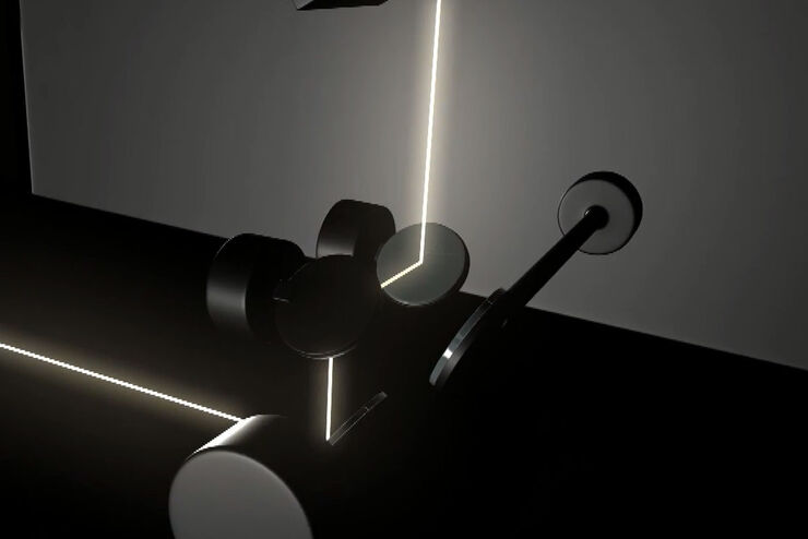
What is a Tandem Scanner?
A Tandem Scanner is an assembly of two different types of scanning together in one system for true confocal point scanning. The Tandem Scanner consists of a three-mirror scanning base with the…
![3D glomeruli in a portion of an ECi-cleared kidney scanned by light sheet microscopy. Courtesy of Prof. Norbert Gretz, Medical Faculty Mannheim, University of Heidelberg [1].](/fileadmin/_processed_/d/d/csm_DLS-Sample-Preparation-Intr_915e0fd7c2.jpg "3D glomeruli in a portion of an ECi-cleared kidney scanned by light sheet microscopy. Courtesy of Prof. Norbert Gretz, Medical Faculty Mannheim, University of Heidelberg [1].")
Using Mounting Frames for Light Sheet Sample Preparation
Sample handling is an important topic in the context of Light Sheet Microscopy. The TCS SP8 DLS integrates Light Sheet technology into an inverted confocal platform and can hence make use of general…

Using a Rotation Device for Light Sheet Sample Mounting
The TCS SP8 DLS from Leica Microsystems is an innovative concept to integrate the Light Sheet Microscopy technology into the confocal microscope. Due to its unique optical architecture samples can be…
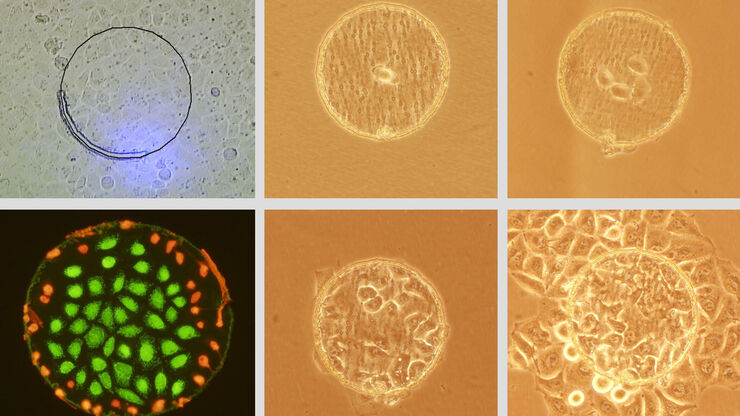
Live Cell Isolation by Laser Microdissection
Laser microdissection is a tool for the isolation of homogenous cell populations from their native niches in tissues to downstream molecular assays. Beside its routine use for fixed tissue sections,…
