Gold EM grid with a silicon dioxide (SiO₂) support film, containing vitrified T47D cells intrinsically expressing GFP-H2B (labeling nuclei) and RFP-LifeAct (labeling the actin cytoskeleton) and high-pressure frozen C. elegans germline tissue, fluorescently labeled with GFP-tagged synaptonemal complex protein

Flujos de trabajo de preparación de muestras EM & Usos

Los investigadores pueden obtener resultados de alta calidad, precisos y reproducibles al obtener imágenes de muestras con microscopía electrónica utilizando las propuestas de preparación de muestras de Leica. Nuestras soluciones admiten microscopía electrónica de barrido (SEM), microscopía electrónica de transmisión (TEM) y criomicroscopía electrónica.

No tiene más que ponerse en contacto con nosotros.

Nuestros expertos en la materia están a su disposición para asesorarle sobre soluciones para flujos de trabajo de preparación de muestras EM & Uses.

¿Cómo pueden los investigadores preparar las muestras para el SEM?

La preparación de muestras para SEM (microscopía electrónica de barrido) implica técnicas como el recubrimiento, el secado y la inclusión en resina para garantizar una imagen óptima. Utilizando las soluciones de Leica para muestras de SEM, los investigadores mejoran la conductividad, reducen los artefactos y garantizan superficies estables y de alta calidad para obtener imágenes de SEM reproducibles a temperatura ambiente.

¿Qué métodos son los mejores para la preparación de muestras de TEM?

La preparación de muestras para el TEM (microscopía electrónica de transmisión) requiere cortes ultrafinos y sin artefactos, recubrimientos de rejillas de TEM y tinción para lograr una visualización detallada de las estructuras. Con la precisión y fiabilidad de los instrumentos de Leica, los investigadores pueden obtener imágenes TEM de alta resolución.

¿Qué es importante para la preparación de muestras criogénicas?

Cryo EM se beneficia de la vitrificación o crío-fijación rápida para mantener las estructuras en su estado nativo.

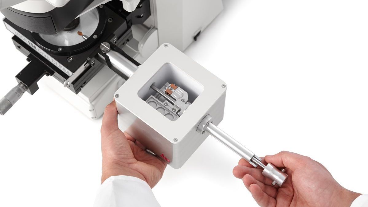
Las soluciones de criopreparación de Leica -incluida la vitrificación avanzada, el recubrimiento, el corte ultrafino, el aplanado criogénico y los flujos de trabajo personalizables de criotransferencia y correlativa - proporcionan muestras reproducibles y libres de contaminación. Ayudan a mantener las estructuras nativas, proporcionan una orientación precisa y permiten la obtención de imágenes criogénicas de alta resolución.

¿Por qué elegir instrumentos y soluciones de Leica para la preparación de muestras EM?


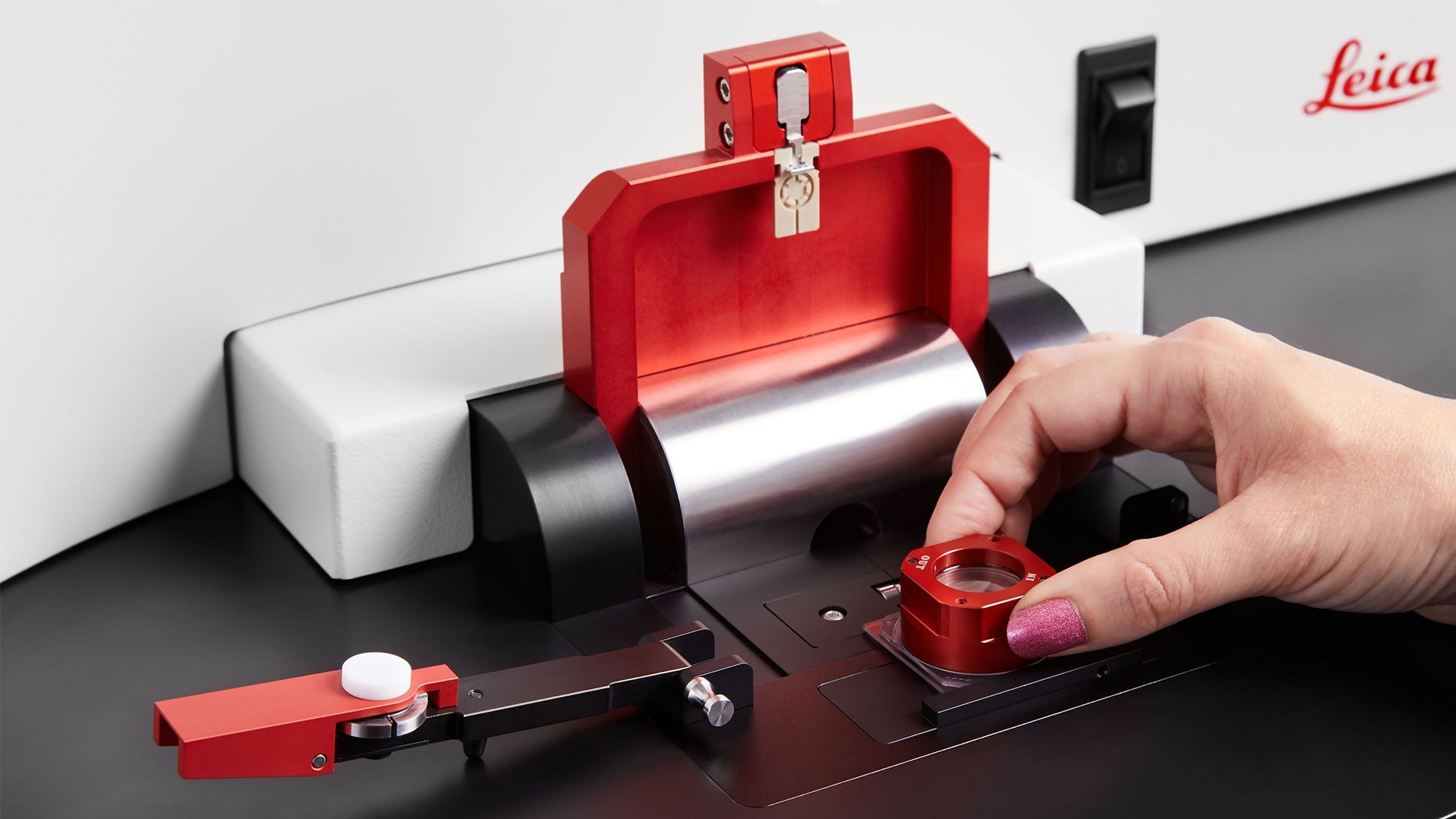
Preparación de muestras de SEM

Las herramientas de preparación de SEM de Leica Microsystems incluyen recubridores sputter de alto vacío y secadores de punto crítico. Estas soluciones garantizan una imagen de las superficies de alta calidad. Además, los instrumentos Leica proporcionan resultados consistentes y fiables para sus estudios de SEM.


Preparación de muestras de TEM

Los investigadores pueden confiar en las soluciones de preparación de TEM de Leica Microsystems por su precisión y fiabilidad. Herramientas esenciales como los ultramicrotomos y los sistemas automatizados de tinción ayudan a producir cortes ultrafinos e imágenes de alta resolución. El recubrimiento por pulverización catódica mejora la conductividad de la muestra y la calidad de la imagen.


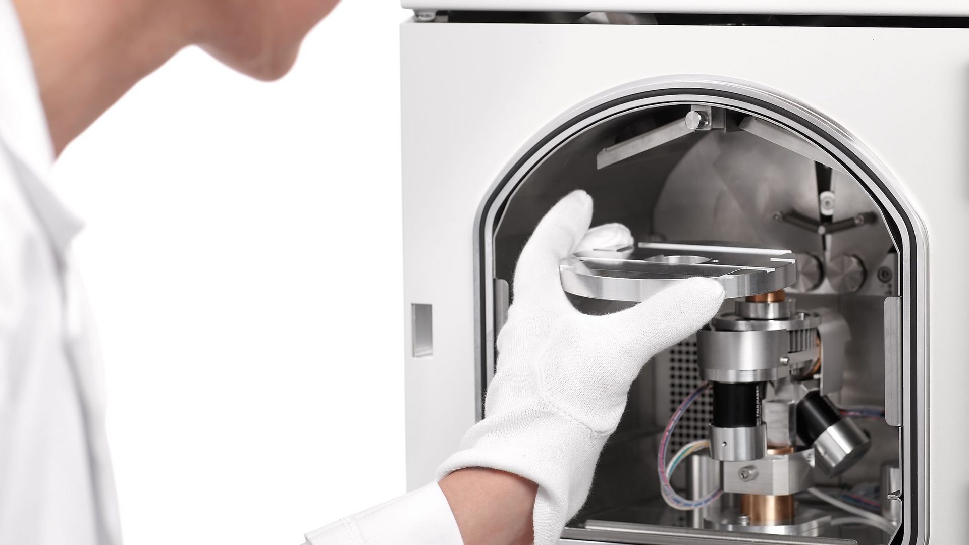
Preparación completa de Cryo-EM

Con los instrumentos de criopreparación de Leica Microsystems, incluidos los vitrificadores por inmersión y los vitrificadores de alta presión, los investigadores garantizan una congelación y vitrificación rápidas. Además, nuestro sistema de transferencia de muestras para flujos de trabajo criogénicos ofrece opciones de transferencia para todo tipo de muestras. Esta opción las protege de la contaminación, garantizando un análisis crio-EM preciso y detallado.
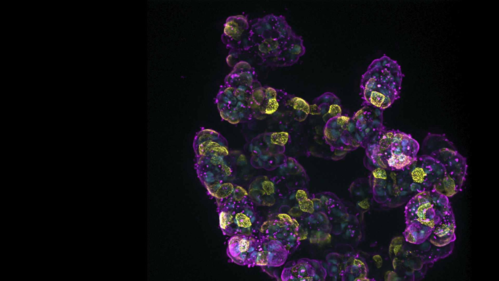
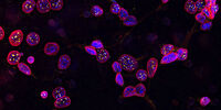
Artículos relacionados

Lea nuestros últimos artículos sobre flujos de trabajo de preparación de muestras EM & Usos

El portal de conocimiento de Leica Microsystems ofrece investigaciones científicas y materiales de aprendizaje sobre microscopía. El contenido está diseñado para ayudar a los investigadores noveles, así como a médicos y científicos experimentados en su trabajo diario o con sus experimentos.

Más artículos
Sectioning resin-embedded sambles

Ultramicrotomy eBook: Targeting, Trimming & Alignment

Ultramicrotomy is evolving rapidly, and today’s microscopes demand high‑quality sections, precise targeting, and reproducible workflows. This eBook brings together expert application notes, automated…
Foraminifera (Ammonia confertitesta) 
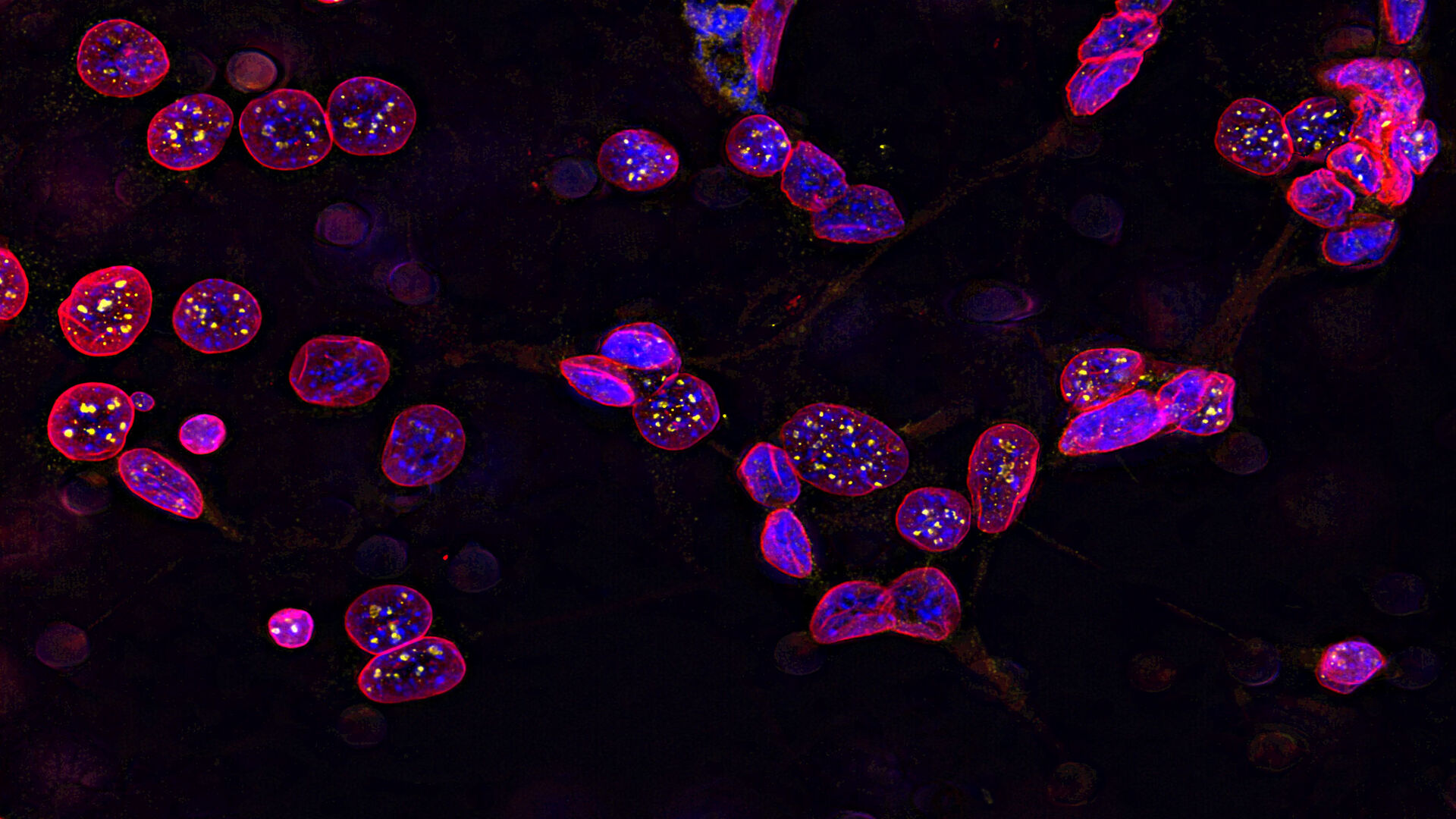
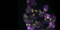
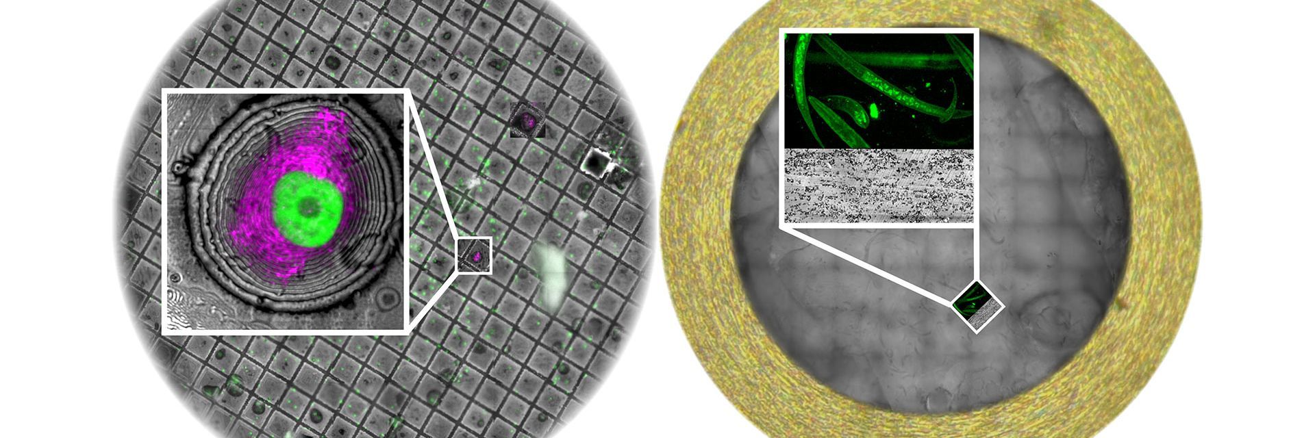
labeled with membrane-permeable calcein, high-pressure frozen in salt water using EM ICE. The sample was cryo-planed and targeted with the M205 on the Cryo-Fluo Enuity, then transferred under cryo conditions to the Cryo-Stellaris for widefield and confocal imaging, revealing details of the staining pattern. Image courtesy: David Evans, University of Southampton.

High-Pressure Freezing for Organoids: Cryo CLEM & FIB Lift Out

Master cryo EM workflow steps for challenging 3D samples: when to choose HPF vs. plunge freezing, reproducible blotting/ice control, contamination aware transfers, Cryo CLEM 3D targeting in organoids,…
Electron microscope (EM) 
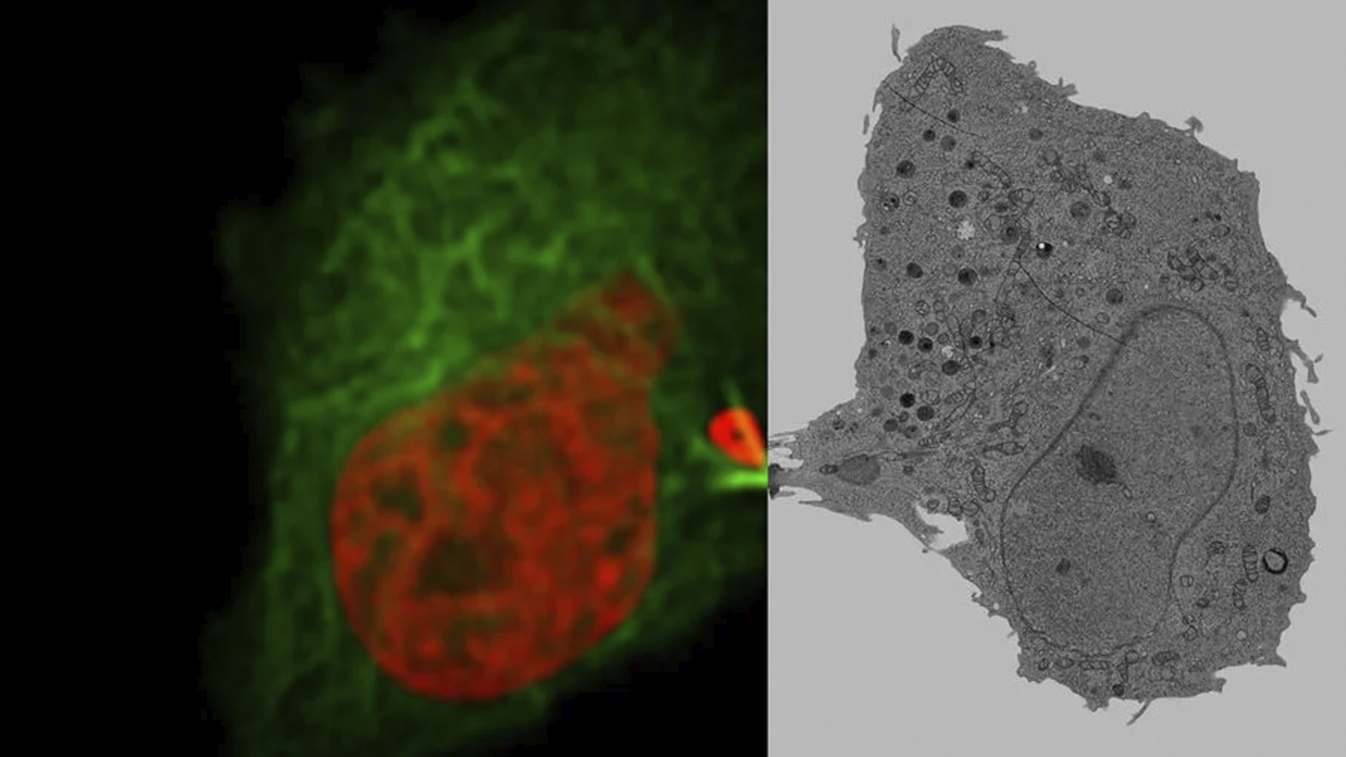
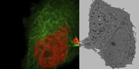
image of a cross section of C. elegans (roundworm). Courtesy of T. Müller-Reichert, MPI-CBG, Dresden, Germany and K. McDonald, University of California, Berkeley, USA.

Brief Introduction to High-Pressure Freezing for Cryo-Fixation

Preparation of biological specimens for electron microscopy (EM) often requires cryo-fixation which does not introduce significant structural alterations of cellular constituents. A common method used…
TEM micrographs of polymer sections. Left: Poly(styrene)-
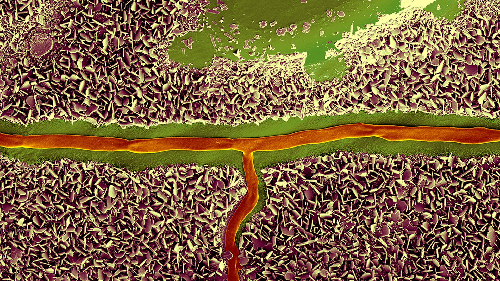
b-poly(isoprene). Right: Poly(styrene)-b-poly(methyl methacrylate).

Ultramicrotome Sectioning of Polymers for TEM Analysis

We demonstrate the capabilities of the UC Enuity ultramicrotome from Leica Microsystems for preparing ultrathin sections of polymer samples under both ambient and cryogenic conditions. By presenting…
Final Segmentation of organelles in Trichomonas species. Magenta – costa, light blue – hydrogenosomes, turquoise – ER, red – vacuoles, yellow – axostyle, green – Golgi apparatus.  Sample courtesy of Isabelle Guerin-Bonne, Low Kay En, Electron Microscopy Unit, Yong Loo Lin School of Medicine, National University of Singapore. Scale bar: 1 µm.

Volume EM and AI Image Analysis

The article outlines a detailed workflow for studying biological tissues in three dimensions using volume-scanning electron microscopy (volume-SEM) combined with AI-assisted image analysis. The focus…

Integrated Serial Sectioning and Cryo-EM Workflows for 3D Biological Imaging

This on-demand webinar explores how integrated tools can support electron microscopy workflows from sample preparation to image analysis. Experts Andreia Pinto, Adrian Boey, and Hoyin Lai present the…
SEM image of the full Li-NMC electrode sample, showing the two porous layers and the metal film at the center of the structure.

Revealing Sodium Battery Degradation via Cryo-EM and CryoFIB

Explore how cryogenic electron microscopy and focused ion beam techniques uncover the intrinsic structure of sodium battery interfaces. This webinar presents a new degradation model based on separator…
Mouse hippocampus brain slice on a grid after HPF using the “Waffle Method”.

The “Waffle Method”: High-Pressure Freeze Complex Samples

This article describes the advantages of a special high pressure freezing method, the so-called “Waffle Method”. Learn how the “Waffle Method” uses EM grids as spacers for high-pressure freezing,…
UC Enuity with Diatome diamond knife.

Mastering Polymer Sectioning with Helmut Gnaegi

When it comes to ultramicrotomy, few names carry the weight of Helmut Gnaegi. As co-founder of Diatome, a global leader in diamond knife technology, Helmut has spent decades refining the art and…
C. elegans embedded in Lowicryl® HM20; pharynx showing red fluorescence (mCherry). The overview shows a front view onto the resin capsule formed by the bottom of a flow-through chamber of the EM AFS2. The capsule was pretrimmed manually. The blockface was trimmed automatically using the AutoTrim function of UC Enuity guided by fluorescence of the worm. Edge length of both squares in relation to the images is 250 µm.

How Fluorescence Guides Sectioning of Resin-embedded EM Samples

Electron microscopes, including transmission electron microscopes (TEM) and scanning electron microscopes (SEM), are widely utilized to gain detailed structural information about biological samples or…
14: 7-day old 3D cell culture of MDCK cells stably transfected with; Mx1-GFP (green), SPY555-Actin (yellow) and WGA-AlexaTM 647 (magenta), and imaged using a THUNDER Imager Cell Spinning Disk. Shown are both the spinning disk image (left) and the same image after THUNDER Computational Clearing was applied (right); scale bar 50 μm.

Organoides y cultivos celulares en 3D

Uno de los avances recientes más emocionantes en la investigación de las ciencias biológicas es el desarrollo de sistemas de cultivo celular en 3D, como organoides, esferoides y modelos de órganos en chip. Un cultivo celular en 3D es un ambiente artificial en el cual las células pueden crecer e interactuar con su entorno en las tres dimensiones. Estas condiciones son similares a un estado in vivo.

Leer más

Image of C2C12 cells: The cells are stained with lamin B (magenta) which indicates nuclear structure, Hoechst (blue) indicating DNA, and γH2AX (yellow) indicating damage to DNA. Cells were imaged using a THUNDER Imager 3D Live Cell with a 63X/1.4 oil immersion objective.

Investigación en biología celular

La obtención de imágenes para biología celular se lleva a cabo con diversos microscopios ópticos y de electrones. Las soluciones para obtener imágenes ofrecidas por Leica Microsystems están diseñadas para maximizar su investigación en biología celular.

Leer más

Mammalian cells expressing H2B-mCherry and alphaTubulin-mEGFP to visualize DNA and microtubules respectively. Left: Widefield imaging using THUNDER Imager. Right: Sample after correlated EM imaging using Leica Microsystems Coral Life workflow. Cells were cryo-immobilized by high-pressure freezing 60 min after the onset of cytokinesis. The intercellular bridge was imaged under TEM after freeze substitution and resin infiltration.

Microscopía correlativa óptica y electrónica (CLEM)

Las soluciones CLEM de Leica Microsystems garantizan la viabilidad de las muestras, la calidad de las comprobaciones y un mecanismo de localización 3D preciso y fiable. Los usuarios pueden utilizar estas soluciones para identificar directamente la célula adecuada en el momento adecuado, obtener imágenes confocales en crio de alta resolución o colocar la información de fluorescencia en el contexto ultraestructural.

Leer más

Cultivo celular

Cultivar células en condiciones de laboratorio es la base del trabajo de los científicos en campos como la biología celular, la investigación del cáncer, la biología del desarrollo y cualquier otra especialidad en el ámbito de las ciencias de la vida y la investigación farmacológica. Descubra cómo Leica ayuda a cultivar células animales en el laboratorio.

Leer más

14: 7-day old 3D cell culture of MDCK cells stably transfected with; Mx1-GFP (green), SPY555-Actin (yellow) and WGA-AlexaTM 647 (magenta), and imaged using a THUNDER Imager Cell Spinning Disk. Shown are both the spinning disk image (left) and the same image after THUNDER Computational Clearing was applied (right); scale bar 50 μm.
Image of C2C12 cells: The cells are stained with lamin B (magenta) which indicates nuclear structure, Hoechst (blue) indicating DNA, and γH2AX (yellow) indicating damage to DNA. Cells were imaged using a THUNDER Imager 3D Live Cell with a 63X/1.4 oil immersion objective.
Mammalian cells expressing H2B-mCherry and alphaTubulin-mEGFP to visualize DNA and microtubules respectively. Left: Widefield imaging using THUNDER Imager. Right: Sample after correlated EM imaging using Leica Microsystems Coral Life workflow. Cells were cryo-immobilized by high-pressure freezing 60 min after the onset of cytokinesis. The intercellular bridge was imaged under TEM after freeze substitution and resin infiltration.
Scroll to top