Produits

Produits

Produits

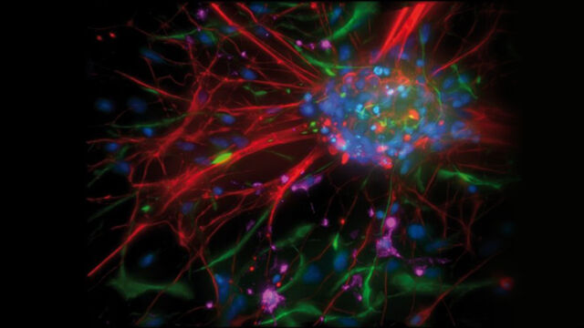
Nous développons nos systèmes de précision haute-technologie pour l'analyse des microstructures avec l'utilisateur, pour l'utilisateur. Dans notre portefeuille de produits, vous trouverez des solutions système pour la science de la vie, comprenant la biotechnologie et la médecine, ainsi que pour la recherche et le développement des matériaux bruts et l'assurance qualité industrielle.

Produits

Nos Dernières Innovations

Consultez le programme de nos prochaines conférences, congrès, salons professionels, webinaires et ateliers et joignez-vous à nous lors d'un de nos prochains événements!

Tous les événements

Lire nos derniers articles

Le portail de connaissances de Leica Microsystems propose des recherches scientifiques et du matériel d'enseignement sur le thème de la microscopie. Le contenu est conçu pour aider les débutants, les praticiens expérimentés et les scientifiques dans leur travail quotidien et leurs expériences.

Plus d'articles

The Guide to Augmented Reality in Microsurgery

In an era of technological advancement, Augmented Reality (AR) is rapidly transforming the medical field. In surgical microscopy, AR can display fluorescence signals as digital overlays in real-time within the surgical field of view, providing surgeons with additional information about fluorescent areas of interest. A surgeon's view can be augmented with data overlays from different systems, offering functional or anatomical information superimposed onto the live microscope view.

How Fluorescence Guides Sectioning of Resin-embedded EM Samples

Electron microscopes, including transmission electron microscopes (TEM) and scanning electron microscopes (SEM), are widely utilized to gain detailed structural information about biological samples or non-living materials. Ultramicrotomy is the preferred technique for producing ultrathin sections, less than 100 nm thick for TEM/SEM analysis. During sample preparation small sample pieces are embedded in epoxy or acrylic resin, excess resin is trimmed away, and the specimen is sliced into ultrathin sections (50 nm - 100 nm) using a glass or diamond knife.

Coherent Raman Scattering Microscopy Publication List

CRS (Coherent Raman Scattering) microscopy is an umbrella term for label-free methods that image biological structures by exploiting the characteristic, intrinsic vibrational contrast of their molecules. The two most important CRS techniques are Coherent Anti-Stokes Raman Scattering (CARS) and Stimulated Raman Scattering (SRS). The biochemical image contrast of CRS is in many ways complementary to the molecular contrast obtained in fluorescence microscopy. A second crucial advantage of these methods is that they preserve the specimen/sample in a near pristine state. This reference list presents current and basic papers on CRS microscopy.

Microscopes de dissection

Si vous devez procéder à des dissections, il peut vous arriver de rester penché sur les oculaires du microscope pendant des heures. Leica Microsystems vous propose de faire votre choix parmi un large éventail de microscopes et une gamme exhaustive de pièces et d'accessoires propres aux microscopes de dissection afin que vous trouviez la solution répondant parfaitement à vos besoins.

A Guide to Polarized Light Microscopy

Polarized light microscopy (POL) enhances contrast in birefringent materials and is used in geology, biology, and materials science to study minerals, crystals, fibers, and plant cell walls.

Factors to Consider when Selecting Clinical Microscopes

What matters if you would like to purchase a clinical microscope? Learn how to arrive at the best buying decision from our Science Lab Article.

Clinical Microscopy: Considerations on Camera Selection

The need for images in pathology laboratories has significantly increased over the past few years, be it in histopathology, cytology, hematology, clinical microbiology, or other applications. They serve many purposes on top of the documentation of the diagnosis. Yet, the view through the eyepieces and the image are different in nature, the one is an optical image, the other a digital image. Looking at a few aspects of this process that are related cameras will help you make sure you can obtain the images with all the detail and color fidelity you need.

Explore Alzheimer's Spatial Proteome with Big Data

Alzheimer's disease, a genetic and sporadic neurodegenerative condition, leads to cognitive decline in mid to late life, marked by β-amyloid plaques and tau tangles. With limited treatment options, new investigative strategies are crucial. The Cell DIVE multiplexed imaging solution allows examination of Alzheimer's brain tissue, potentially uncovering new research avenues. Here, we showcase the Cell DIVE image viewer, enabling users to access the full Alzheimer's multiplexed dataset directly in their browser.
Scroll to top