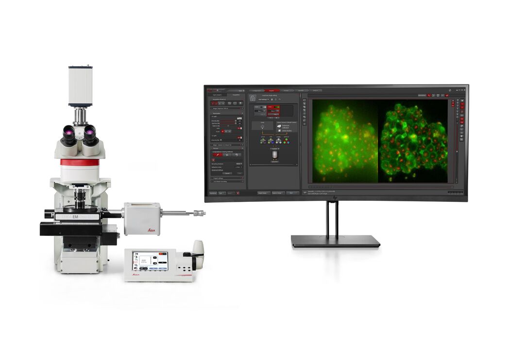
THUNDER Imager EM Cryo CLEM Cryomicroscope optique

Une compréhension approfondie de la biologie structurelle cellulaire

Lire nos derniers articles

How to Successfully Perform Live-cell CLEM

The Leica Nano workflow provides a streamlined live-cell CLEM solution for getting insight bout structural changes of cellular components over time. Besides the technical handling described in the…

How to Successfully Implement Coral Life

The live-cell  CLEM workflow allows you to capture dynamic information related to a relevant biological process as it happens and put these observations into their ultrastructural context. The Leica…

Advancing Cellular Ultrastructure Research

Freeze-fracture and freeze-etching are useful tools for studying flexible membrane-associated structures such as tight junctions or the enteric glycocalyx. Freeze-fracture and etching are two…

The Cryo-CLEM Journey

This article describes the Cryo-CLEM technology and the benefits it can provide for scientists. Additionally, some scientific publications are highlighted. Recent developments in cryo electron…
3D reconstruction of an intercellular bridge in a C. elegans embryo

Download EM Workflow Solutions Booklet

This publication is a compilation of appropriate workflows for the most frequently used sample preparation methods, like Correlative Methodologies, Optogenetics & Electro-Physiology, Surface Analysis,…

Exploring the Structure and Life Cycle of Viruses

The SARS-CoV-2 outbreak started in late December 2019 and has since reached a global pandemic, leading to a worldwide battle against COVID-19. The ever-evolving electron microscopy methods offer a…
Cryo FIB lamella - Overlay of SEM and confocal fluorescence image. Target structure in yeast cells (nuclear pore proteine Nup159-Atg8-split Venus, red) marked by an arrow. Scale bar: 5 µm. Alegretti et al.,  Nature 586, 796-800 (2020).

Targeting Active Recycling Nuclear Pore Complexes using Cryo Confocal Microscopy

In this article, how cryo light microscopy and, in particular cryo confocal microscopy, is used to improve the reliability of cryo EM workflows is described. The quality of the EM grids and samples is…

Advancing Cell Biology with Cryo-Correlative Microscopy

Correlative light and electron microscopy (CLEM) advances biological discoveries by merging different microscopes and imaging modalities to study systems in 4D. Combining fluorescence microscopy with…

Workflows and Instrumentation for Cryo-electron Microscopy

Cryo-electron microscopy is an increasingly popular modality to study the structures of macromolecular complexes and has enabled numerous new insights in cell biology. In recent years, cryo-electron…
Mouse retina was fixed and stained by following reagents: anti-CD31 antibody (green): Endothelia cells, IsoB4 (red): Blood vessels, and microglia anti-GFAP antibody (blue): Astrocytes Sample courtesy by Jeremy Burton, PhD and Jiyeon Lee, PhD, Genentech Inc., South San Francisco, USA. Imaged by Olga Davydenko, PhD (Leica). Imaged with a THUNDER Imager 3D Cell Culture.

An Introduction to Computational Clearing

Many software packages include background subtraction algorithms to enhance the contrast of features in the image by reducing background noise. The most common methods used to remove background noise…

Crystal Clear Cryo Light-microscopy Images

This article describes how computational clearing of cryo light microscopy images improves the identification of cellular targets for cryo electron-microscopy.

Real Time Images of 3D Specimens with Sharp Contrast Free of Haze

THUNDER Imagers deliver in real time images of 3D specimens with sharp contrast, free of the haze or out-of-focus blur typical of widefield systems. They can even image clearly places deep inside a…

Domaines d'application

Microscopie Fluorescente

La fluorescence est l'un des phénomènes physiques les plus couramment utilisés en microscopie biologique et analytique, principalement en raison de sa grande sensibilité et de sa haute spécificité.…

Recherche sur le cancer

Le cancer est une maladie complexe et hétérogène causée par des cellules déficientes dans la régulation de la croissance. Les modifications génétiques et épigénétiques d’une cellule ou d’un groupe de…

Biologie cellulaire

Découvrez comment vous pouvez tirer le meilleur parti de vos recherches en biologie cellulaire et élargir vos découvertes grâce aux solutions d’imagerie de Leica Microsystems.

Virologie

Vos recherches portent sur les infections et les maladies virales ? Découvrez comment renforcer vos connaissances en virologie grâce aux solutions d’imagerie et de préparation d’échantillons de Leica…

Organismes modèles dans la recherche

Un organisme modèle désigne une espèce utilisée par les chercheurs pour étudier des processus biologiques spécifiques. Ils présentent des caractéristiques génétiques similaires à celles de l’homme et…

Tomographie cryoélectronique

La tomographie cryoélectronique (CryoET) est utilisée pour analyser les biomolécules dans leur environnement cellulaire avec une résolution sans précédent, inférieure au nanomètre.

Techniques avancées de microscopie

Les techniques de microscopie avancées englobent les techniques d'imagerie à haute résolution et à super-résolution. Ces techniques sont principalement utilisées pour visualiser des événements…

Microscopie corrélative CLEM

Les workflows Coral de Leica Microsystems aident les utilisateurs à corréler les données entre la microscopie de fluorescence et la microscopie électronique (CLEM).
Background image
Scroll to top