Aivia AI Image Analysis Software
A complete analysis workflow from accurate deep-learning based cell segmentation to automatic phenotyping and data exploration for 3D multiplexed images.
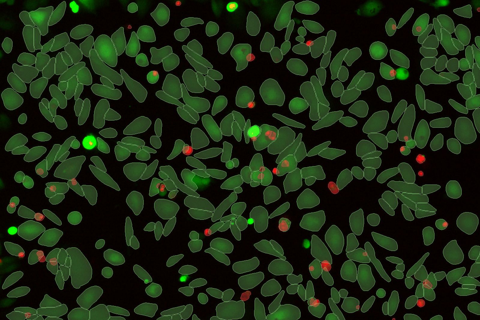
Subjectivity of analysis and poor reproducibility are key hurdles to be overcome for biological image analysis. Standard segmentation can lead to sub-standard results and require substantial manual curation which is subject to human error. Aivia changes all of that.
Grafici interattivi per dati relativi a più pozzetti
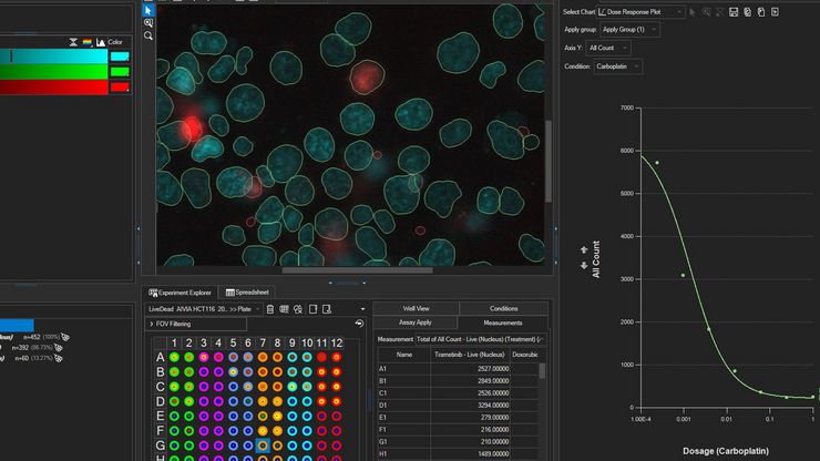
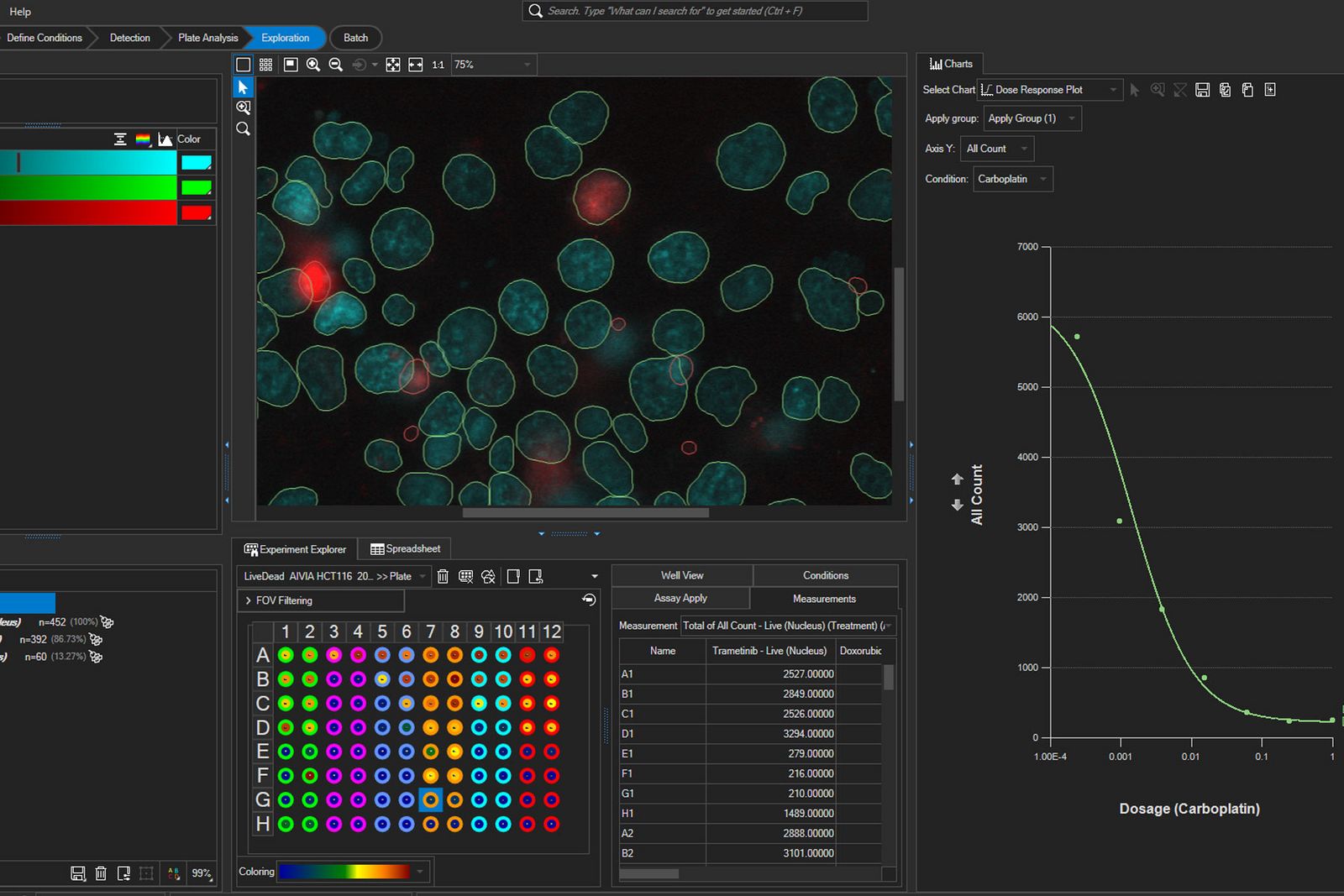
Esplorazione avanzata dei dati per esperimenti su più pozzetti. Aivia 16 ottimizza l'analisi dei dati e consente di ottenere informazioni più approfondite sugli esperimenti multi-pozzetto grazie a grafici riassuntivi aggregati e all'esportazione dei dati per l'analisi statistica.
- Estrarre informazioni approfondite da set di dati aggregati relativi a più pozzetti, per singolo pozzo, per insieme di oggetti o per condizione
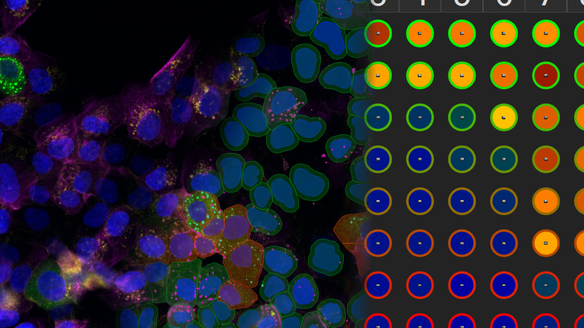
- Riassumi e esplora i risultati delle analisi per ciascun pozzetto grazie alla mappa termica multipozzetto nell'Experiment Explorer
- Personalizza l'aggregazione dei dati nei grafici di riepilogo
- Scopri la risposta cellulare in un esperimento a pozzetti multipli a partire dalla curva dose-risposta con il calcolo di IC50/EC50
- Esporta i dati aggregati in un software di analisi statistica di terze parti per approfondire l'analisi dei risultati
AI access for all
Aivia makes advanced data analysis accessible for all biologists - with no computer science expertise required.
The Aivia platform has been designed with the end-user in mind. This means with Aivia you can quickly and reliably generate high-quality results. The Aivia platform includes all state-of-the-art applications you will need in a unified user experience.
Quickly train laboratory users on the platform, to conduct their analysis without any specialist expertise.
- Speed up your imaging projects and publish faster
- Benefit from next-generation, easy to use machine learning segmentation and classification tools
- Conduct parameter-free image segmentation
- Easily train, update and apply deep learning models using local resources or the AiviaCloud platform
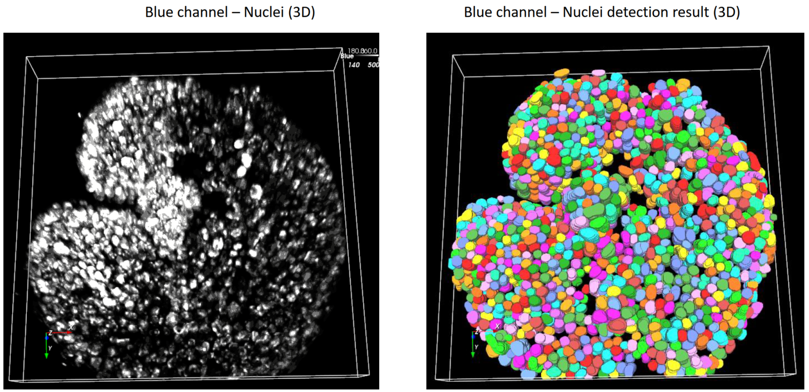
Total freedom on a single platform
Aivia's powerful and fast 2-5D visualization and analysis unlocks all the value of your data - within a single platform.
No longer does your team have to learn to operate and adopt multiple imaging and analyses systems into their workflow - the Aivia platform unifies all state-of-the-art applications you will need in a unified user experience. Aivia can leverage both local and cloud computing resources. You can install and use Aivia both on your local computer as well as via a web browser, AiviaWeb. Aivia works seamlessly with all microscopy imaging systems.
Your team can also access all files created by your imaging systems anywhere - all you need is an internet connection.
- Powerful and fast 2-5D visualization and analysis - accessible anywhere
- Includes 22 applications and 20 pre-trained deep learning models (image segmentation, restoration and virtual staining)
- Reliable and easy to use cloud access with flexible IT architectures supported
- Over 45 microscopy file formats supported

Start a free trial
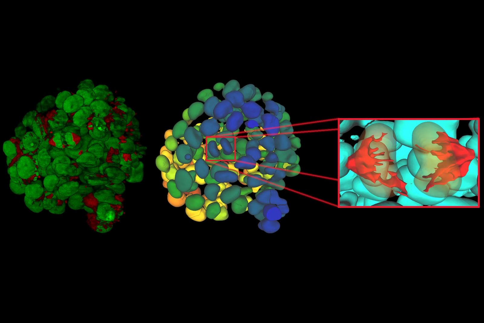
Using state-of-the-art, AI-first software architecture, Aivia is a uniquely innovative and complete 2-to-5D image visualization, analysis and interpretation platform designed for the reliable processing and reconstruction of highly complex images in just minutes.
- Make AI-enhanced image analysis accessible for all - with no computer science expertise required
- Leverage machine learning capabilities to generate robust and reproducible segmentation results
- Realize powerful and fast 2-5D visualization and analysis to unleash the value of your data - all within a single platform
Explore Aivia subscription plans
Aivia is available via subscription, giving you the flexibility to select a plan that fits your lab’s needs now, or in the future as your research needs evolve.
With technical support and free software upgrades for the duration of your subscription, you will always have the lasted AI-powered analysis tool for your research.
- Go - The essential tools to get you started with AI image analysis
- Elevate - Take your AI image analysis to the next level with CellBio or Neuro
- Apex - A comprehensive AI image analysis platform for labs and core facilities
Everything you need to start analyzing images
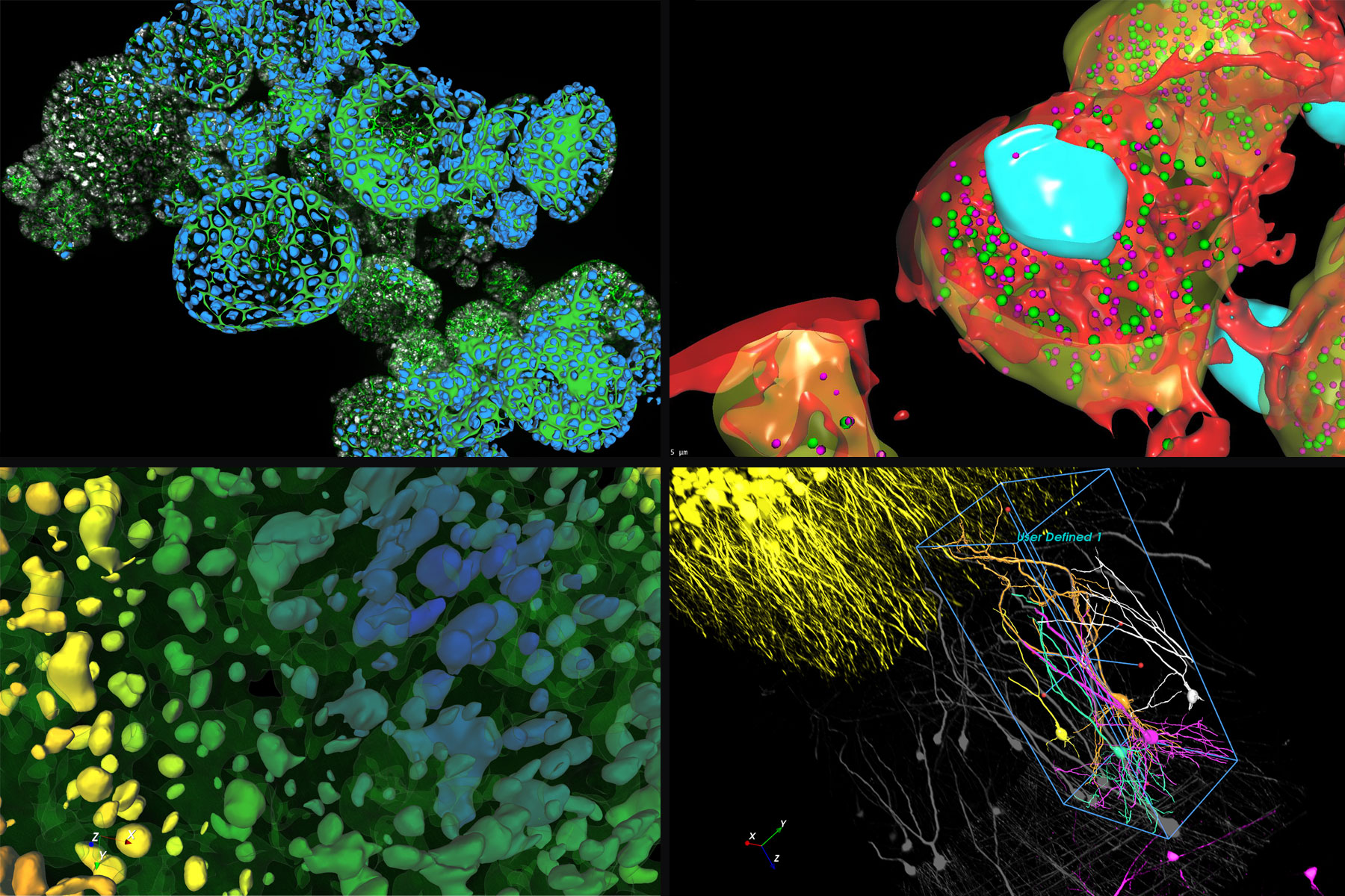
Aivia Go offers state-of-the-art image visualization and analysis tools in a single platform, including multiple AI-powered features, to meet your challenging image visualization and analysis needs. Simple segmentation workflows and batch processing capabilities yield results faster, helping you leap from data to publication.
Aivia Go supports a large range of applications from 2D to 5D image analysis tasks such as detection and tracking. It is the ideal package for research laboratories and imaging core facilities. No computer expertise required.
- AI-powered classifiers
- 12+ image analysis recipes
- 2D -5D image visualization
Features
Image Analysis Recipes - Deploy a wide range of 2D to 5D image analysis recipes for the most popular analysis applications:
- Count cells, nuclei and particles in 2D and 3D
- Track cells (in phase contrast and fluorescence), nuclei, and particles in 2D and 3D
- Cell proliferation and wound healing assays (phase contrast)
- Neurite outgrowth
- Stem cell colony detection (phase contrast)
- Cell tracking (phase contrast)
Descubrimiento rápido de información en experimentos multipocillos complejos
Aivia Accelerate permite una rápida exploración de las relaciones entre variables clave en experimentos multipocillos complejos, permitiendo a los científicos extraer información procesable de los datos de imagen con una configuración mínima y sin necesidad de codificación.
Características
- Analice los resultados agregados en todas las condiciones experimentales utilizando gráficos de resumen con estadísticas de población
- Acelere los análisis con flujos de trabajo preconfigurados
- Despliegue de un algoritmo generalista de segmentación de células aprendizaje profundo preentrenado para la segmentación por ejemplos
- No se requiere entrenamiento de modelos, codificación ni experiencia en IA
- Clasifique automáticamente las poblaciones celulares
- Generación automática de informes exhaustivos
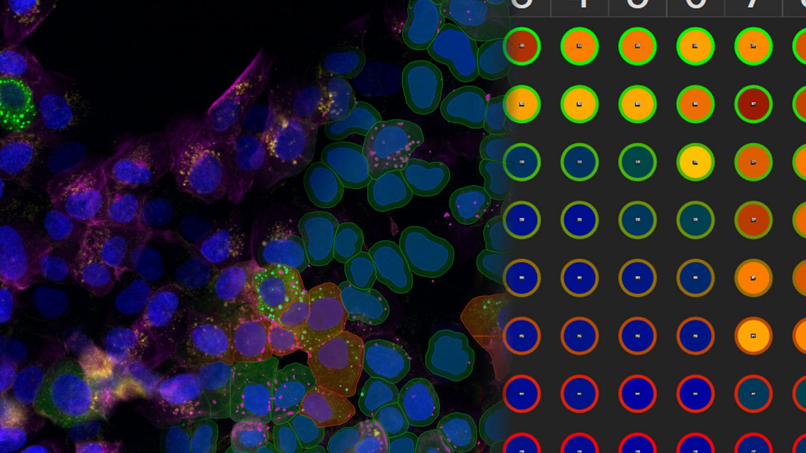
Take your AI image analysis to the next level with Aivia’s simple workflows
A complete solution for research laboratories and core imaging facilities, Aivia Elevate subscribers can choose specialized AI solutions for either neuro or cell biology image visualization and analysis.
Analyze cellular interactions and measure the distances between different compartments down to the channel level with Elevate for CellBio . It includes tools to perform accurate cell detection in 2D multiplexed images, identify cell phenotypes and explore spatial insights. Or automate challenging neuroimaging tasks, such as neuron tracing, dendrite segmentation, and more with Elevate for Neuro.
Want both options? Upgrade to Aivia Apex for full functionality plus the ability to apply your own deep learning models.
Features
Cell or Neuron Analysis Recipes - Choose between specialized analysis tools for neuroscience or cell biology research
- Neuron Analysis Recipes (Fluorescence/EM): Detect and trace neurons in 3D data sets
- Cell Analysis Recipes (2D/3D): Observe complex cellular interactions in 2D and 3D data sets
A comprehensive AI image analysis platform for labs and core facilities
Aivia Apex is a comprehensive microscopy image analysis solution for researchers who need a variety of image analysis applications. Apex also provides microscopists with the flexibility to apply their own deep learning models, third party models or open-source repositories.
Ideal for large research groups and core imaging facilities, Aivia Apex’s Floating License Manager allows a site to run all licenses on any machine on their local network.
- Everything in Elevate Neuro & CellBio
- Expanded deep learning capabilities
- Floating license manager
Features
Image Analysis Recipes - Deploy a wide range of 2D to 5D image analysis recipes for the most popular analysis applications
- Nuclei count and tracking
- Cell count and tracking
- Particle count and tracking
- Cell proliferation assay
- Neurite outgrowth
- Wound healing (phase contrast)
- Stem cell colony detection (phase contrast)
- Cell tracking (phase contrast)
- 3D Object Analysis and Tracking recipes including neuron and cell analysis
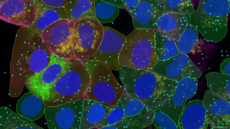
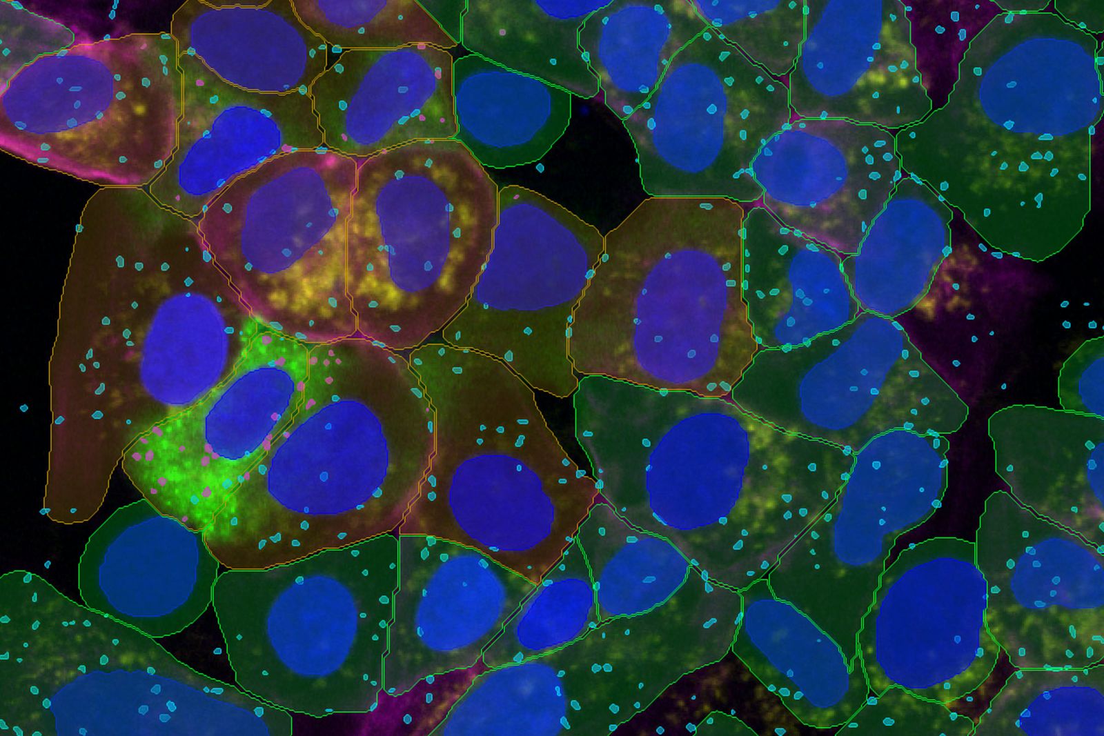
Funzione Segment by example – evoluta
Più approfondimenti, più velocemente e con meno dati da inserire. Segment by Example di Aivia 16 ridefinisce la semplicità e la precisione, sfruttando l'innovativo algoritmo di deep learning di Aivia, in attesa di brevetto, per identificare le cellule con elevata accuratezza e il minimo sforzo.
- Segmenta con precisione le cellule in 2D o 3D con soli tre campioni
- Combina più canali di immagine per il rilevamento di cellule multiplex 2D/3D
- Segmenta cellule, nuclei e un numero illimitato di tipi di vescicole in 2D
- Caratterizza fenotipicamente popolazioni cellulari eterogenee in un'unica operazione
- Rileva cellule e nuclei fino a 12,8 volte più velocemente
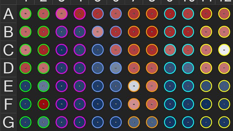
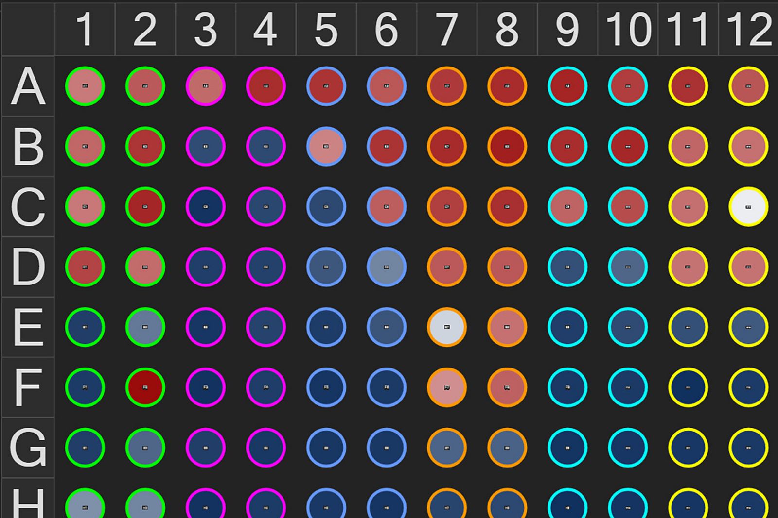
Importazione di dati su più pozzetti
Importazione di dati su più pozzetti senza problemi. Aivia 16 consente ai ricercatori di importare, esplorare e analizzare i dati senza problemi. Importa nuovi esperimenti multi-pozzetto direttamente da file LIF o XELF dai sistemi di imaging Mica, DMi8 e STELLARIS.
Passa con facilità tra un pozzetto all’altro e da una posizione di campo all’altra in un esperimento con più pozzetti ed esamina rapidamente i dati caricati in Aivia grazie al rinnovato Experiment Explorer.
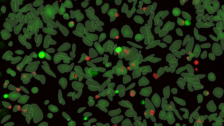
Analisi assistita del saggio
Flussi di lavoro semplificati per l'analisi end-to-end dei saggi. L'analisi guidata semplifica il processo di scoperta, garantendo maggiore accuratezza e affidabilità sui campioni multi-pozzetto e offrendo prestazioni ai vertici della categoria agli utenti che desiderano trarre il massimo valore dai propri dati.
- Apri la sequenza guidata per i saggi da Analysis Launchpad
- Le sequenze guidate accompagnano gli utenti dall'impostazione delle condizioni fino al rilevamento, alla fenotipizzazione e all'elaborazione in batch
- Quattro (4) sequenze guidate predefinite per saggi comuni: conteggio, analisi morfologica, viventi/morti e apoptosi
- Risultati dell'analisi aggregati per pozzetto, per condizione o per piastra
- Ottimizza la pipeline di analisi utilizzando un'immagine di esempio e applicare i risultati all'intero esperimento