
病理学における顕微鏡観察に関する課題は何ですか?
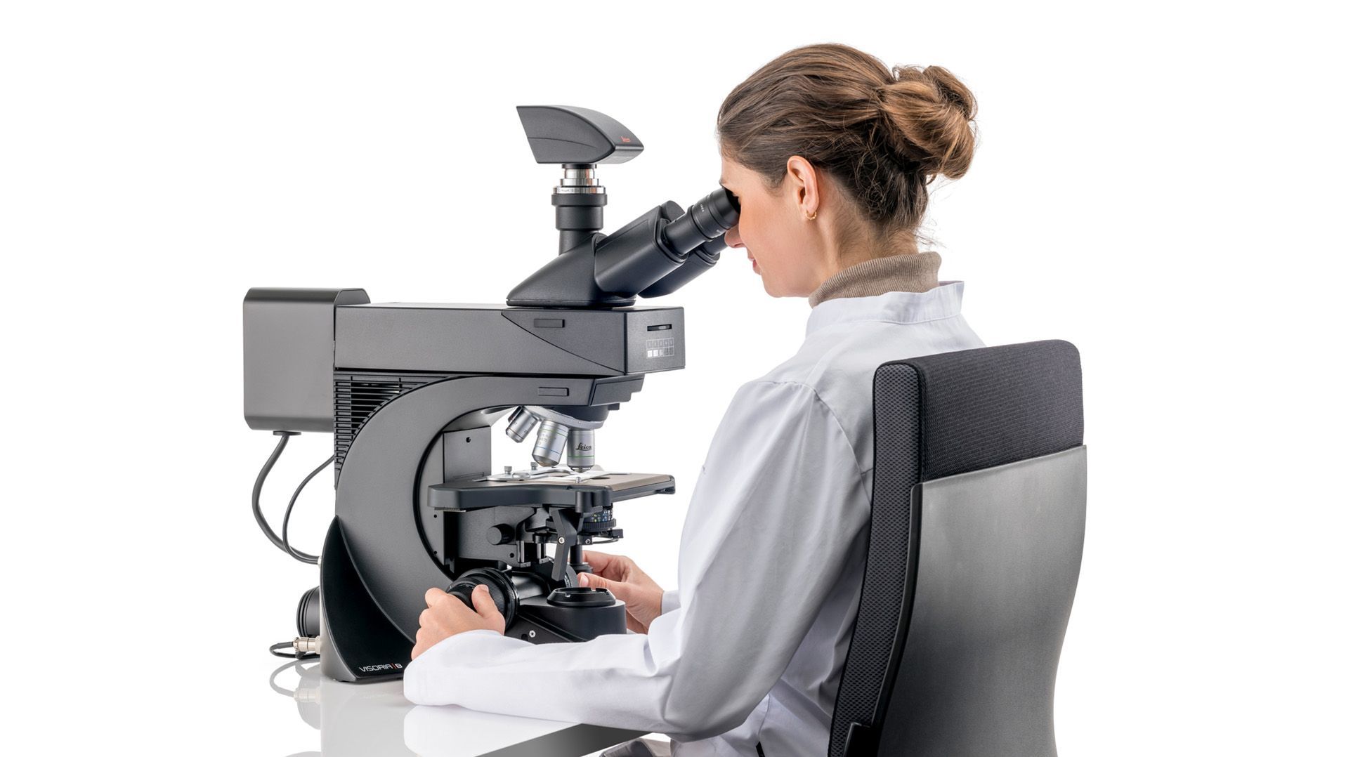
病理医は、顕微鏡観察のために標本スライドを作製し、診断の正確性と信頼性を確保する必要があります。この作業は、病院や検査施設における臨床意思決定を迅速に支援する必要があるため、厳しい時間制約の中で行われることが多いです。ライカの顕微鏡は、病理医が効率的かつ信頼性の高い標本処理を実現するのに役立ちます。

病理学において顕微鏡はどのように使用されますか?
顕微鏡観察とは、病理検査の工程において顕微鏡を用いて実施されるすべての検査を指します。例えば、標本の染色品質のチェックや、標本の観察および記録(ドキュメンテーション)などが含まれます。Leicaの顕微鏡は、病理診断を効率的かつ快適に行えるようユーザーをサポートします。

なぜ病理学者は顕微鏡を使用するのですか?
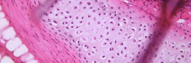
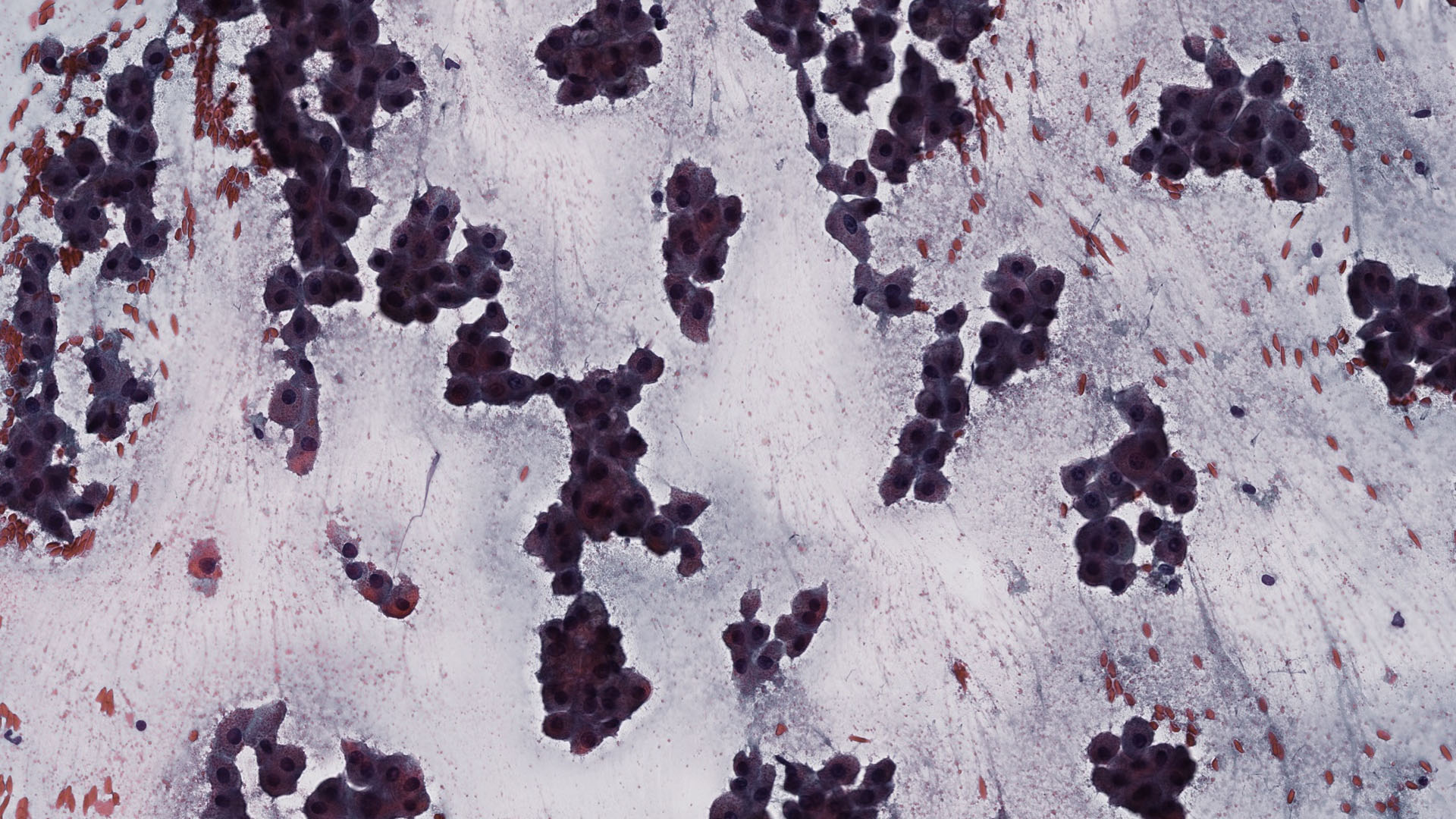
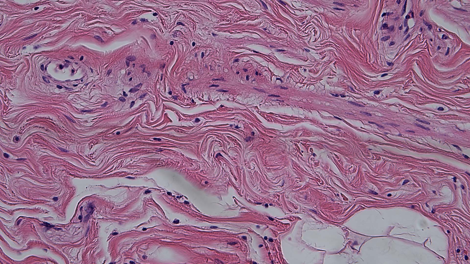
顕微鏡を用いることで、病理医は微細構造や微妙な色の差、さらには標本中の特定細胞の数など、肉眼では把握できない異常を観察することが可能になります。Leicaの顕微鏡は、多様なコントラスト法により、より詳細な構造の観察を可能にします。

臨床病理(Clinical Pathology)と解剖病理(Anatomic Pathology)の違いは何でしょうか?
臨床病理と解剖病理では、対象とする検体が異なります。臨床病理には、臨床化学、分子病理学、免疫病理学、血液病理学、そして医療微生物学が含まれます。解剖病理は、外科病理、細胞診、法医学病理、皮膚病理学などで用いられる、組織や細胞の組織学的(病理学的)検査に焦点を当てています。病理医は、効率的な診断を実現できるライカの顕微鏡によって恩恵を受けています。
臨床検査や病理分野において、なぜLeicaの顕微鏡が選ばれるのでしょうか?

病理学についてよくある質問
カメラはドキュメンテーション、ライブ像での画像表示、カンファレンスなど拡大像を共有して検討等に使用されます。 また、特に画像に注釈を挿入したり、研究所や病院の情報システムにアーカイブすることが可能なソフトウェアを搭載したカメラであれば、結果の報告にも活用できます。
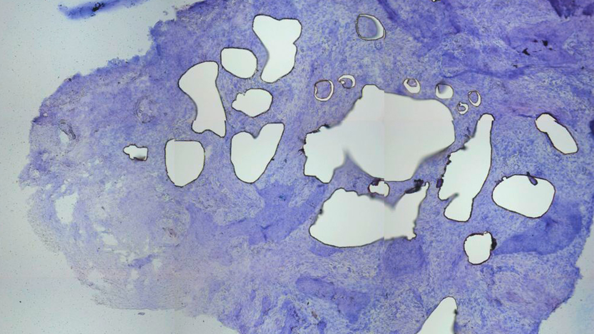
顕微鏡検査では、何を見る必要があるかが重要です。 染色試料の構造や色合いを見る時は、明視野で試料を観察します。 非染色の細胞や組織の構造を特定する必要がある時は、位相差を使用します。 詳しくは、サイエンスラボの記事をご覧ください。 Factors to Consider when Selecting Clinical Microscopes.
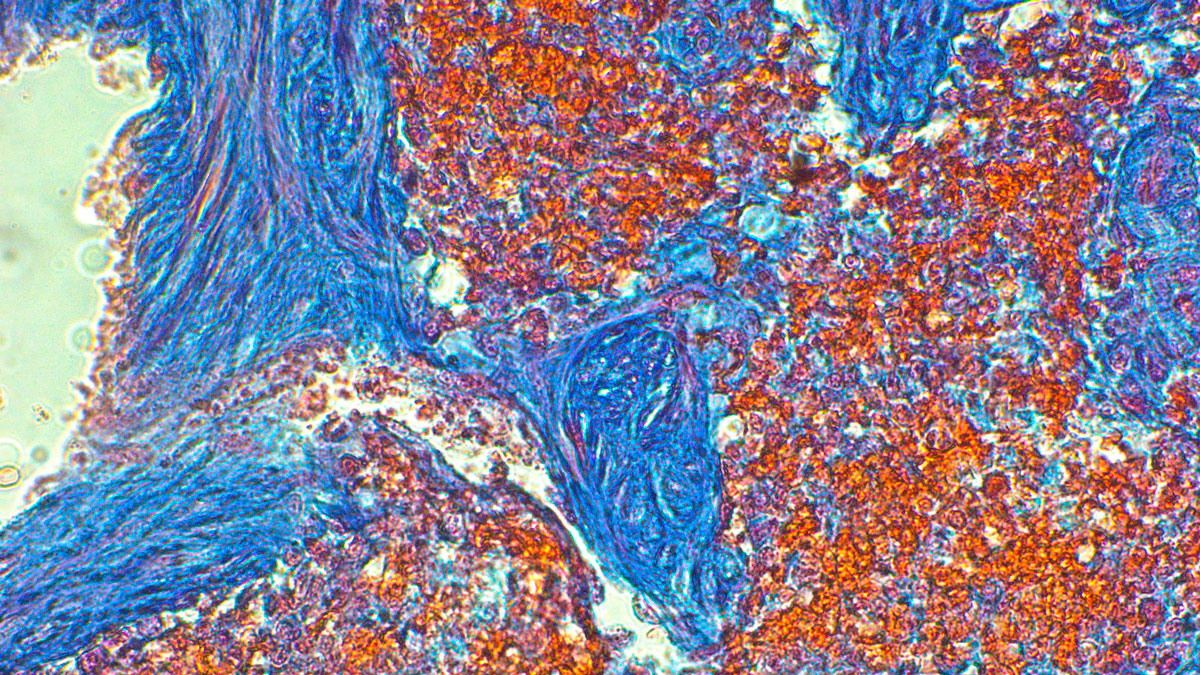
カメラにはカラーとモノクロがあります。 カラーカメラは染色内に存在する微差を視覚化し、試料について豊富な情報をもたらすため、病理学用途に最適です。 モノクロカメラはFISH(蛍光in situハイブリダイゼーション)等の蛍光アプリケーションに最適です。 詳しくは、サイエンスラボの記事をご覧ください。 Clinical Microscopy: Considerations on Camera Selection.
