DM1000とDM3000顕微鏡の比較表
| DM1000 LED | DM2000 & DM2000 LED | DM2500 & DM2500 LED | DM3000 & DM3000 LED | |
| 時々使用 | x | - | - | - |
| 中~高負荷 | - | x | x | x |
| 接眼レンズにより、顕微鏡の高さと視野角の調節が可能 | x | x | x | x |
| 左右対称レイアウトのコントロールノブ | - | x | x | x |
| 調整可能なステージの高さとフォーカスコントロール | x | x | x | x |
| フォーカスノブのトルク調整可能 | - | x | x | x |
| 複数のユーザーが同時に観察できるマルチビューシステム | - | - | x | - |
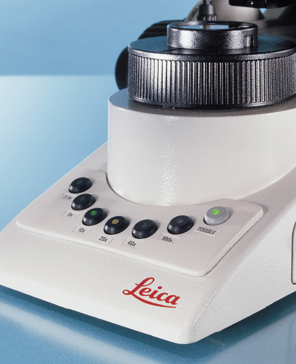
| スイッチングモードでの素早い対物レンズ交換 | - | - | - | x |
| 自動照明管理 | - | - | - | x |
| コンパクトで堅牢な設計 | x | x | x | x |
| 製品ページへ | 製品ページへ | 製品ページへ | 製品ページへ |
x = 含まれる, - = 含まれない
Pathology Solution Suite の利点
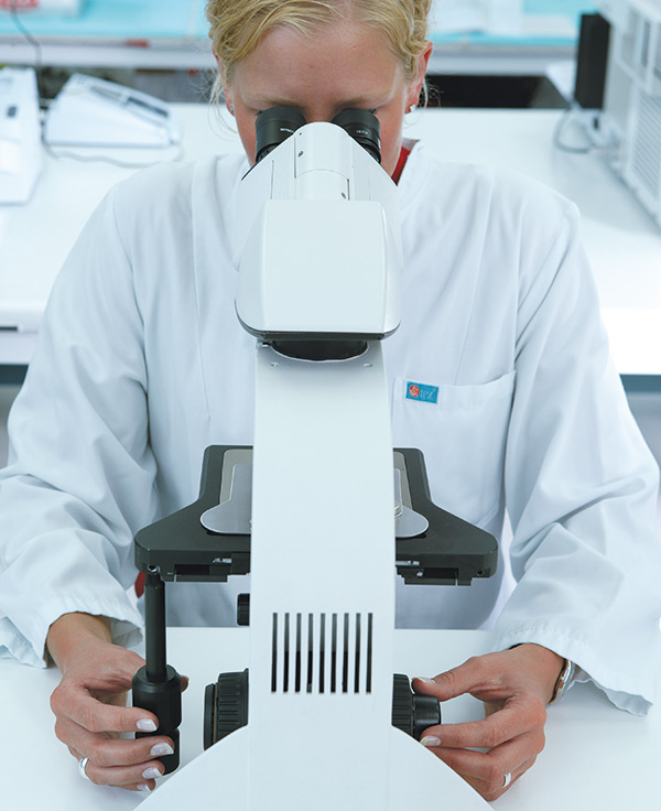
Customer experiences with a DM3000 microscope for clinical applications
Trudi de Jong and Marianne Noordanus, both from Rotterdam, describe how a DM3000 microscope helps them to perform their clinical microscope work more comfortably and efficiently.
Courtesy of:
Trudi de Jong, Erasmus MC academic hospital Rotterdam, the Netherlands, Hematology
Marianne Noordanus, Star-MDC, Medisch Diagnostisch Centrum, Rotterdam (The Netherlands) Microbiology
病理学についてよくある質問
カメラはドキュメンテーション、ライブ像での画像表示、カンファレンスなど拡大像を共有して検討等に使用されます。 また、特に画像に注釈を挿入したり、研究所や病院の情報システムにアーカイブすることが可能なソフトウェアを搭載したカメラであれば、結果の報告にも活用できます。
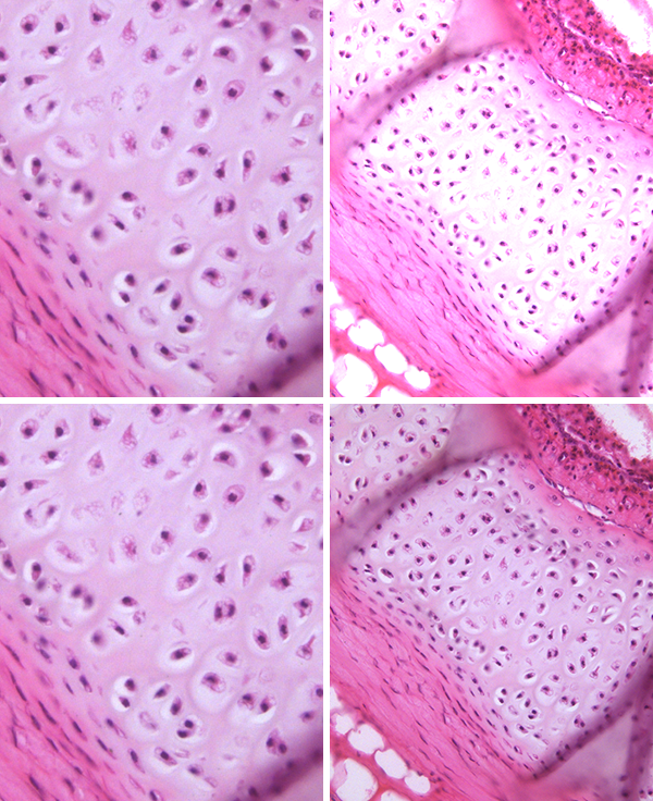
顕微鏡検査では、何を見る必要があるかが重要です。 染色試料の構造や色合いを見る時は、明視野で試料を観察します。 非染色の細胞や組織の構造を特定する必要がある時は、位相差を使用します。 詳しくは、サイエンスラボの記事をご覧ください。 Factors to Consider when Selecting Clinical Microscopes.
カメラにはカラーとモノクロがあります。 カラーカメラは染色内に存在する微差を視覚化し、試料について豊富な情報をもたらすため、病理学用途に最適です。 モノクロカメラはFISH(蛍光in situハイブリダイゼーション)等の蛍光アプリケーションに最適です。 詳しくは、サイエンスラボの記事をご覧ください。 Clinical Microscopy: Considerations on Camera Selection.
