お問い合わせ

Focus on Microscopy 2023

02 Apr 2023 08:30 - 05 Apr 2023 15:30 UTC

Alfândega do Porto Congress Center, R. Nova da Alfândega, 4050-387 Porto, Portugal

Congress

Let’s inspire your research!

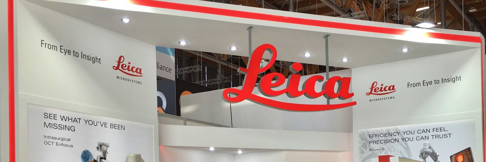
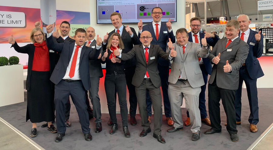
Join us at the FOM 2023 at booth 8-9.

Our talks
Monday, 3 April, 12:20 - 12:40
High content screening of spheroids using light sheet microscopy
A. Diosdi, I. Grexa, A. Sebestyen, P. Horvath, J. Reddington1 (1Leica Microsystems CMS GmbH)
High content screening (HCS) of 3D multicellular spheroids offers an opportunity to generate data with high translatability for drug discovery and drug validation studies.

We present a workflow that combines an automated spheroid picker with a newly developed HCS plate for high content image acquisition using the light sheet microscope of Leica Microsystems (DLS).

Monday, 3 April, 16:10 - 16:30
The more the merrier: Extending confocal microscopy with multicolor unmixing tools for advanced multiplexing and spatial omics
J. Roberti, S. Hoffmann, I. Steinmetz, J. Reddington, L.A.J. Alvarez (Leica Microsystems CMS GmbH)
The need to understand the complex nature of biological processes is constantly driving science to find new and better ways to study them. This need and novel palettes of fluorophores have pushed imaging technologies towards an increasing number of signals that one can probe on a single sample. This opened the door to advanced multiplexing and imaging “omics” approaches, to reveal key aspects of tissue organization, cancer progression, tumor-immune interactions, and infectious diseases.

The STELLARIS confocal platform offers different approaches to achieve multiplexing in a direct straightforward manner. We will demonstrate the acquisition of 11 fluorophores within a sample in one imaging round and discuss how lifetime-based tools in STELLARIS enable going beyond these spectral capabilities. This allows scientific research to extend the possibilities of detecting signals in higher order of multiplexing experiments.

Wednesday, 5 April, 10:20 - 10:40
Artificial intelligence in the microscopy workflow
J. Reymann (Leica Microsystems CMS GmbH)

Artificial Intelligence (AI) tools to simplify daily life tasks, are revolutionizing the world. 

AI tools for image analysis have been developed in order to enable researchers to segment objects which cannot be detected by standard tools, or which could only be detected by manual segmentation or counting. We will discuss how such tools can be incorporated into the microscopy workflow, in order to enable researchers to gather the data they need in the easiest way possible, and to expand the capabilities of state-of-the-art instruments for experiments that were previously not possible due to time and/or complexity.

Book your demo at our booth on

  • Digital Transmitted Light Microscope Mateo TL 
  • Laser Microdissection Microscope LMD
  • Confocal Microscope STELLARIS 5 with White Light Laser and Light Sheet
  • Live-cell CLEM Workflow Coral Life 
  • THUNDER Imager with THUNDER Live
  • AI Image Analysis Software Aivia
Scroll to top